Review
. 2024 Nov 22;13(12):1439.
doi: 10.3390/antiox13121439.

Potential Opportunities for Pharmacogenetic-Based Therapeutic Exploitation of Xanthine Dehydrogenase in Cardiovascular Disease

Affiliations
Review

Potential Opportunities for Pharmacogenetic-Based Therapeutic Exploitation of Xanthine Dehydrogenase in Cardiovascular Disease

Gianmichele Massimo et al. Antioxidants (Basel). .

Abstract

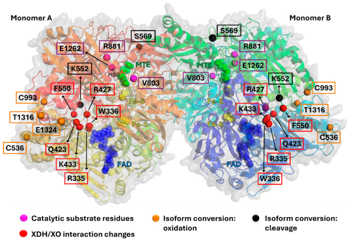
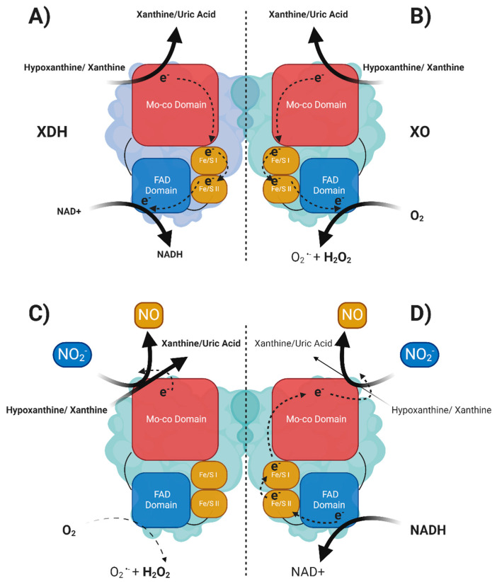
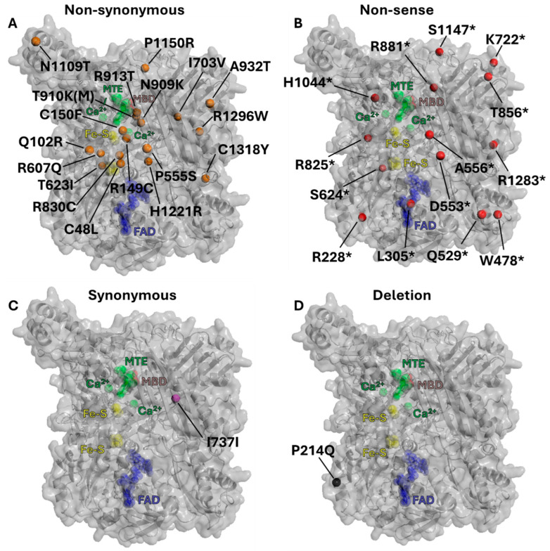
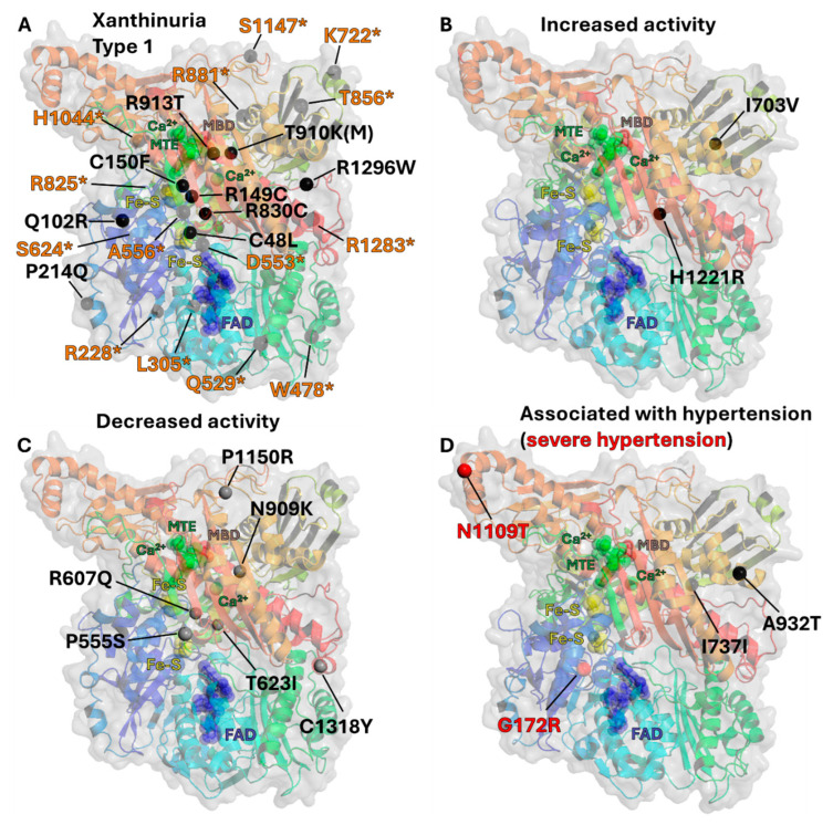
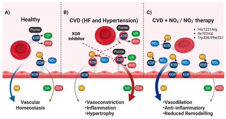
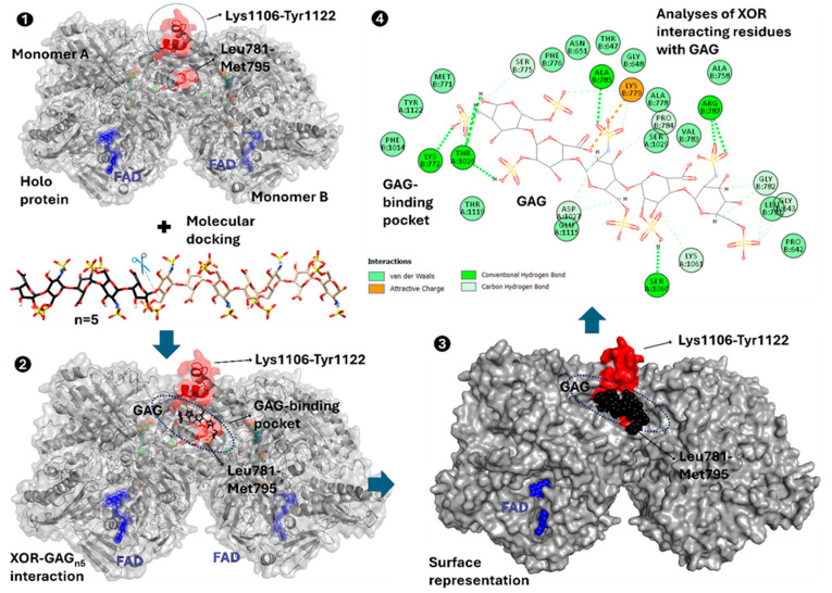
The majority of naturally occurring mutations of the human gene XDH, are associated with reduced or completely absent xanthine oxidoreductase (XOR) activity, leading to a disease known as classical xanthinuria, which is due to the accumulation and excretion of xanthine in urine. Three types of classical xanthinuria have been identified: type I, characterised by XOR deficiency, type II, caused by XOR and aldehyde oxidase (AO) deficiency, and type III due to XOR, AO, and sulphite oxidase (SO) deficiency. Type I and II are considered rare autosomal recessive disorders, a condition where two copies of the mutated gene must be present to develop the disease or trait. In most cases, xanthinuria type I and II result to be asymptomatic, and only occasionally lead to renal failure due to urolithiasis caused by xanthine deposition. However, in the last 10-15 years, new observations have been made about the link between naturally occurring mutations and pathological phenotypes particularly pertinent to cardiovascular diseases (CVD). These links have been attributed to a genetically driven increase of XOR expression and activity that is responsible for what is thought to be damaging uric acid (UA) and reactive oxygen species (ROS) accumulation, nitric oxide (·NO) depletion and endothelial dysfunction. In this review, we discuss the importance of genetics for interindividual variability of XOR expression and activity while focusing mainly on those variants thought to be relevant for CVD. In addition, we discuss the potential exploitation of the genetically driven increase of XOR activity to deliver more beneficial bioavailable ·NO. Finally, we examine the effect that non-synonymous mutations have on the tertiary structure of the protein and consequently on its capacity to interact with glycosaminoglycans (GAGs) localised on the outer surface of endothelial cells.

Keywords: NO; cardiovascular diseases; glycosaminoglycans; nitrite; nitrite reduction; xanthine oxidoreductase.


Conflict of interest statement

The authors declare the following financial interests/personal relationships which may be considered as potential competing interests: Amrita Ahluwalia is a Co-Director of Heartbeet Ltd. and IoNa Therapeutics seeking to identify therapeutic opportunities for dietary nitrate.

Figures

Figure 1
Figure 1
Position of key functional residues in XOR. Purple Residues: Required for functional enzymatic molybdenum dependent activity. Red Residues: interaction between these residues is lost in the XDH to XO isoform conversion. Orange Residues: sites of cystine oxidation, which induces XDH to XO isoform conversion. Black Residues: sites of proteolytic cleavage which induces XDH to XO isoform conversion. Figure derived from human XOR crystal structure (PDB 2E1Q). Also shown are (Green) substrate MTE (phosphonic acidmono-(2-amino-5,6-dimercapto-4-oxo-3,7,8a,9,10,10a-hexahydro-4h-8-oxa-1,3,9,10-tetraaza-anthracen-7-ylmethyl)ester) in the molybdenum active pocket, (Yellow) Fe-S clusters, (Blue) FAD in the FAD domain, and (MBD) molybdenum.
Figure 2
Figure 2
Schematic of XOR homodimer structure and the catalysed reactions and electron flux within an XOR monomer. (A) XOR monomer in the XDH state oxidises purines at the Mo-co domain. The electrons at the reduced Mo-co domain are shuttled to the FAD domain via the Fe/S clusters restoring the Mo-co domain oxidation state (Mo+4 to Mo+6). The XDH isoform has an increased affinity to utilise NAD+ as a terminal electron acceptor. (B) XO has an increased affinity for O2 as a terminal electron acceptor and produces O2•− and H2O2. (C) Electrons donated from purines oxidation are utilised by the Mo-co domain to reduce NO2 which prevents the formation of O2•− and H2O2 at the FAD domain (this reaction is favoured in low O2 concentrations). (D) NADH acts as a reducing substrate and donates electrons at the FAD domain which, via retrograde electron flux, reduces the Mo-co domain. The Mo-co domain can reduce NO2 to NO, which resolves its oxidation state. This NADH reduced Mo-co domain favours NO2 and reduces UA production (this reaction favours high NADH concentrations and acidic pH).
Figure 3
Figure 3
Position of residue mutations which attenuate XOR activity. (A) Orange residues indicate sites of non-synonymous mutations. (B) Red residues indicated by * represent sites of non-sense mutations that also mediate Xanthinuria Type 1 as presented in Table 1. (C) Purple residue indicates a site of synonymous mutation. (D) Black residue indicates a site of deletion mutation. Figure derived from human XOR crystal structure (PDB 2E1Q). Also shown are (Green) substrate MTE in the molybdenum active pocket, (Yellow) Fe-S clusters, (Blue) FAD in the FAD domain, and (Brown) molybdenum (MBD).
Figure 4
Figure 4
Position of residue mutations which induce a phenotype. (A) Residues indicate sites of non-synonymous and non-sense mutations associated with Xanthinuria Type 1. * highlights non-sense mutation site residues (Orange) as presented in Table 1 (B) Residues indicate sites of non-synonymous mutations associated with increased XOR activity. (C) Residues (Gray) indicate sites of non-synonymous mutations associated with decreased XOR activity. (D) Residues indicate sites of non-synonymous and synonymous mutations that are associated with (Black) hypertension, or (Red) severe hypertension. Figure derived from human XOR crystal structure (PDB 2E1Q). Also shown are (Green) substrate MTE in the molybdenum active pocket, (Yellow) Fe-S clusters, (Blue) FAD in the FAD domain, and (Brown) molybdenum (MBD).
Figure 5
Figure 5
Vascular localisation of XOR in health and disease. (A) Healthy individuals have modest XOR expression in the vasculature which is either extracellularly bound to glycosaminoglycans (GAGs) in the vessel lumen, or intracellularly expressed in endothelial cells. Healthy individuals have a relative balance between XOR NO2 reductase capacity and production of UA and ROS which contribute to vascular homeostasis. (B) In CVD, upregulated expression and activity of RBC and endothelial XOR results in enhanced UA and ROS production. The increased production of UA and ROS contribute to attenuate NO signalling and promotes vasoconstriction, inflammation, and cardiac hypertrophy. CVD patients with XOR mutations that impair NO2 reductase activity may not experience increased CVD risk with febuxostat administration. In conjunction, they will not be able to utilise NO3/NO2 intervention and may not have any benefit from the repurposing of the enzyme. (C) The elevated XOR expression and activity in CVD patients could alternatively be exploited, via NO3 and NO2 therapy. Increased levels of exogenously derived NO2 could restore the attenuated NO signalling and reduce UA and ROS production. This intervention could be more beneficial in patients that harbour mutants (e.g., His1221Arg, and Ile703Val) with enhanced NO2 reductase capacity. This could convert mutants that may have greater propensity to generate ROS and UA from pathogenic to disease resolving. Overall, this could then help stratify CVD patients for which XOR intervention may be appropriate—inhibition or exploitation ensuring reduced risk with XOR inhibitors or efficacy with dietary NO3 intervention.
Figure 6
Figure 6
Preliminary in silico docking experiments of XOR–GAG interactions. Workflow of experimental approach: (1) Highlighting areas of XOR residues (Leu781-Met795 and Lys1106-Tyr1122) and areas that have been associated with GAG interactions for docking of a small heparin structure (PDB:1HPN was truncated to n = 5 residues); (2) The results of preliminary docking experiments of the heparin (black) fragment into regions of interest (red); (3) Demonstrates the surface representation of the heparin fragment docking into a well-defined interface pocket; (4) Residues Lys772, Ser775, Lys779, Leu781, Gly782, Pro784, Ala785, Arg787, Thr1026, Asp1027, Ser1060, Lys1061, Glu1115, Thr1119, and Tyr1122 form strong electrostatic interactions with the sulfonate groups on heparin. Therefore, any genetic mutations that have the capacity to interfere with these key residues could impact the binding, stability, and affinity of GAGs to XOR. Colored Blue is FAD in the FAD domain.

Similar articles

References

    1. Battelli M.G., Bolognesi A., Polito L. Pathophysiology of circulating xanthine oxidoreductase: New emerging roles for a multi-tasking enzyme. Biochim. Biophys. Acta (BBA)-Mol. Basis Dis. 2014;1842:1502–1517. doi: 10.1016/j.bbadis.2014.05.022. - DOI - PubMed
    1. Roussos G.G. Studies on a hypoxanthine oxidase from bovine small intestine. Biochim. Biophys. Acta. 1963;73:338–340. doi: 10.1016/0926-6569(63)90170-6. - DOI - PubMed
    1. Waud W.R., Rajagopalan K.V. Purification and properties of the NAD+-dependent (type D) and O2-dependent (type O) forms of rat liver xanthine dehydrogenase. Arch. Biochem. Biophys. 1976;172:354–364. doi: 10.1016/0003-9861(76)90087-4. - DOI - PubMed
    1. Jarasch E.D., Bruder G., Keenan T.W., Franke W.W. Redox constituents in milk fat globule membranes and rough endoplasmic reticulum from lactating mammary gland. J. Cell Biol. 1977;73:223–241. doi: 10.1083/jcb.73.1.223. - DOI - PMC - PubMed
    1. Jarasch E.D., Grund C., Bruder G., Heid H.W., Keenan T.W., Franke W.W. Localization of xanthine oxidase in mammary-gland epithelium and capillary endothelium. Cell. 1981;25:67–82. doi: 10.1016/0092-8674(81)90232-4. - DOI - PubMed

LinkOut - more resources