. 2024 Nov 28;13(12):1459.
doi: 10.3390/antiox13121459.

Exploring Oxidative Stress and Metabolic Dysregulation in Lung Tissues of Offspring Rats Exposed to Prenatal Polystyrene Microplastics: Effects of Melatonin Treatment

Affiliations

Exploring Oxidative Stress and Metabolic Dysregulation in Lung Tissues of Offspring Rats Exposed to Prenatal Polystyrene Microplastics: Effects of Melatonin Treatment

Hong-Ren Yu et al. Antioxidants (Basel). .

Abstract

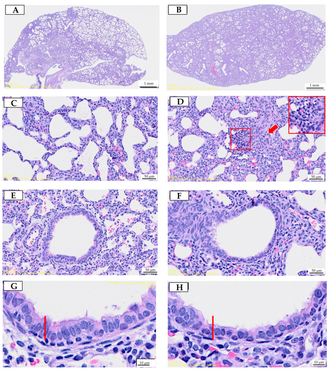
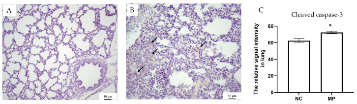
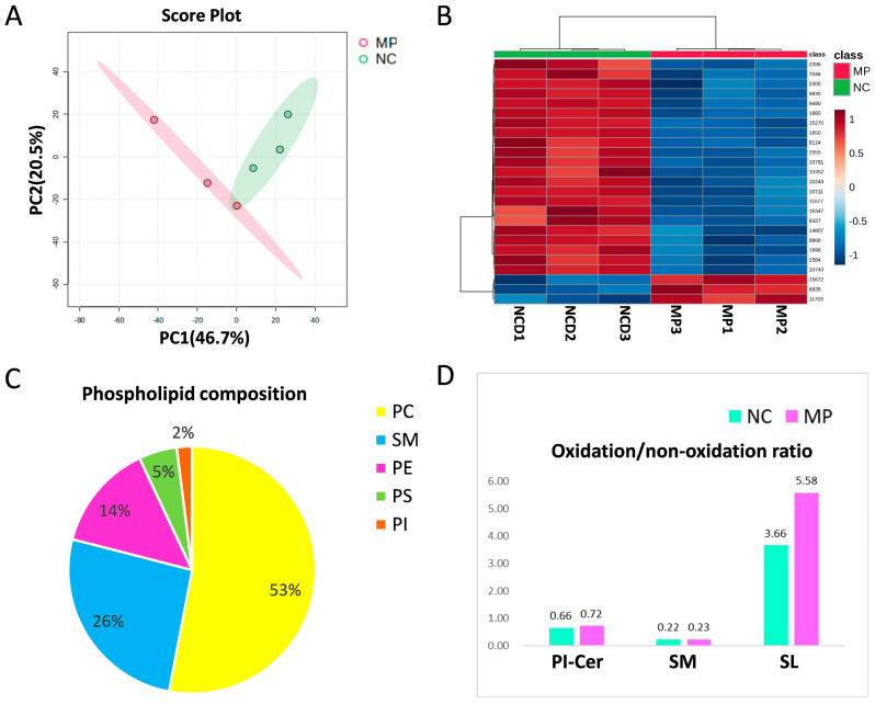
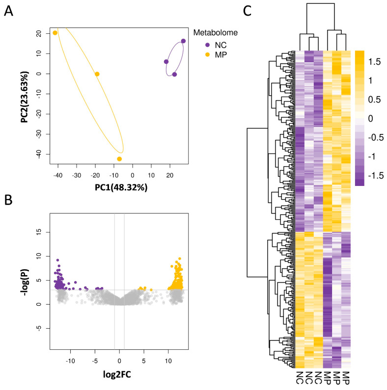
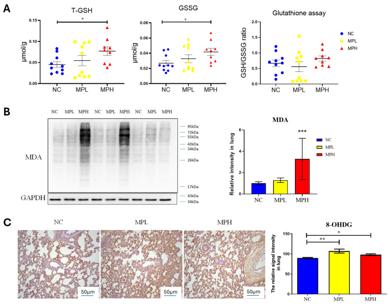
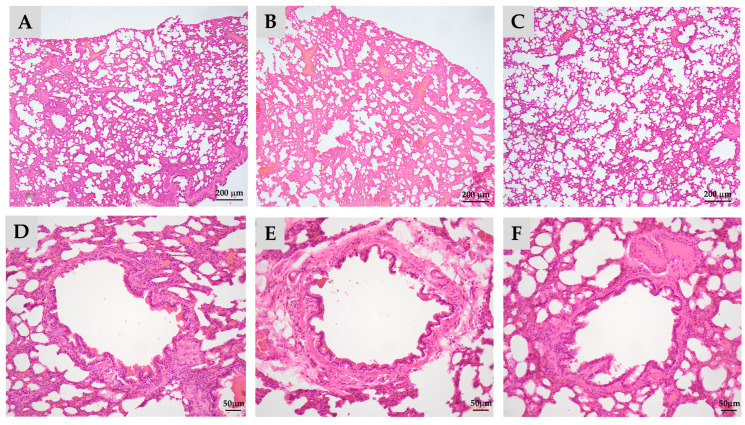
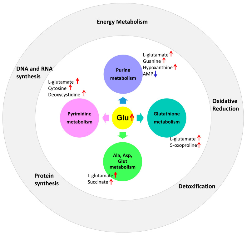
Metabolomics research provides a clearer understanding of an organism's metabolic state and enables a more accurate representation of its functional performance. This study aimed to investigate changes in the metabolome of lung tissues resulting from prenatal exposure to polystyrene microplastics (PS-MPs) and to understand the underlying mechanisms of lung damage in rat offspring. We conducted metabolomic analyses of lung tissue from seven-day-old rat pups exposed to prenatal PS-MPs. Our findings revealed that prenatal exposure to PS-MPs led to significantly increased oxidative stress in lung tissues, characterized by notable imbalances in nucleic acid metabolism and altered profiles of specific amino acids. Furthermore, we evaluated the therapeutic effects of melatonin treatment on lung function in 120-day-old offspring and found that melatonin treatment significantly improved lung function and histologic change in the affected offspring. This study provides valuable biological insights into the mechanisms underlying lung damage caused by prenatal PS-MPs exposure. Future studies should focus on validating the results of animal experiments in humans, exploring additional therapeutic mechanisms of melatonin, and developing suitable protocols for clinical use.

Keywords: lung; melatonin; metabolome; microplastics; offspring; oxidative stress.


Conflict of interest statement

The authors declare no conflicts of interest. The funders had no role in the design of the study; in the collection, analyses, or interpretation of data; in the writing of the manuscript; or in the decision to publish the results.

Figures

Figure 1
Figure 1
Prenatal polystyrene microplastics (PS-MPs) exposure leads to lung dysplasia in offspring during infancy. Histological manifestations of lung tissue from seven-day-old offspring with maternal microplastic particle exposure are shown. (A) Control group: under low-power microscopic field, the control group exhibited better pulmonary tissue aeration development. (B) PS-MPs exposure (1000 µg/L in drinking water for pregnant dams). (C) Zoomed-in images of Figure 1A. (D) Zoomed-in images of Figure 1B, showing the collapsed alveoli and thickened alveolar septa (red arrow). Magnification of boxed area detailing the inflammatory cells; cross-section of small airway from the (E) control group and (F) prenatal PS-MPs exposure group. (G) Zoomed-in images of (E). (H) Zoomed-in images of (F). The columnar epithelial cells in the airway of the prenatal PS-MPs exposure group are shorter and less developed, as indicated by the red marker representing the height of the control group cells. Each group comprised six animals. The yellow label at the bottom left of (F) and the top right of (G) is a specimen group annotation, with no other significance.
Figure 2
Figure 2
Cleaved caspase-3 staining for (A) the control and (B) PS-MPs group. The staining of cleaved caspase-3 is indicated with arrows. (C) Cleaved caspase-3 densities were quantified and compared between different groups. NC denotes the control group, while MP indicates PS-MPs exposure during pregnancy. Each group comprised six animals. Significant difference is indicated by *: p < 0.05.
Figure 3
Figure 3
The differentially expressed lipid-soluble metabolites between the control group (NC) and prenatal PS-MPs exposure group (MP). (A) Principal component analysis (PCA) and (B) heatmap for lipid-soluble metabolites. The heatmap illustrates the top 25 metabolites with the most prominent differences between the two groups. In this display, the rows represent metabolite codes, columns represent samples, and the color intensity within each cell signifies the level of abundance (red = high; blue = low). (C) The composition of phospholipid from lung tissue of seven-day-old rat offspring. (D) Oxidized PI-Cer and SM in control group and in prenatal PS-MPs exposure group. Each group comprised three animals. Abbreviations: Cer, ceramide; PC, phosphatidylcholine; PE, phosphatidylethanolamine; PI, phosphatidylinositol; PS, phosphatidylserine; PA, phosphatidic acid; PG, phosphatidylglycerol; SM, sphingomyelin.
Figure 4
Figure 4
Prenatal exposure to PS-MPs altered the aqueous metabolite composition of offspring lung tissue. (A) The PCA displays the expression of aqueous metabolites in the control group (NC) and the prenatal polystyrene microplastics (PS-MPs) exposure group (MP). The yellow and purple dots represent three samples from the control group and the PS-MPs group, respectively. (B) The volcano plot demonstrates the differential expression of aqueous metabolites between the MPs and control groups. In this study, differential expression analysis of aqueous metabolites was performed, and a threshold of p < 0.05 and |fold change| ≥ 2 was used to identify significant differential metabolites. In the plot, the horizontal axis represents the fold change, while the vertical axis represents the adjusted p-value, with smaller values indicating more significant differences. Higher −log10 values correspond to higher statistical significance. (C) The heatmap shows the differential aqueous metabolites between the MPs group and control group. Increased aqueous metabolites were represented in yellow, and decreased aqueous metabolites were represented in purple, compared to the control group. Each group comprised three animals.
Figure 5
Figure 5
Oxidative stress in the lung tissue of pups. (A) Total glutathione (T-GSH), oxidized glutathione (GSSG), and ratio of reduced glutathione (GSH) to GSSG. Each group comprised 9–10 animals. (B) Western blot of Malondialdehyde (MDA) expression and semi-quantitative analysis. (C) 8-hydroxy-2-deoxyguanosine (8-OHdG) staining and semi-quantitative analysis. Magnification at 40× *: p < 0.05; **: p < 0.01; ***: p < 0.001. Each group comprised six animals.
Figure 6
Figure 6
Melatonin treatment partially ameliorates pulmonary hypoplasia in adult offspring caused by prenatal and postnatal exposure to polystyrene microplastics (PS-MPs). Histological manifestations of lung tissue from 120-day-old offspring under low-power microscopic field. (A) Control group; (B) prenatal plus postnatal PS-MPs exposure group showing the alveolar collapse and hypertrophied alveolar septa; (C) prenatal plus postnatal PS-MPs exposure with melatonin treatment group; (D) zoomed-in images of (A); (E) zoomed-in images of (B) showing thickened connective tissue beneath the epithelial layer; (F) zoomed-in images of (C). Each group comprised three animals.
Figure 7
Figure 7
Prenatal microplastic exposure affects the metabolic pathways of purine, pyrimidine, alanine/aspartate/glutamate, and glutathione in the lung tissue of the offspring, along with their corresponding biological functions. The red upward arrow represents an increase, while the blue downward arrow represents a decrease.

Similar articles

Cited by

References

    1. Cowger W., Willis K.A., Bullock S., Conlon K., Emmanuel J., Erdle L.M., Eriksen M., Farrelly T.A., Hardesty B.D., Kerge K., et al. Global producer responsibility for plastic pollution. Sci. Adv. 2024;10:eadj8275. doi: 10.1126/sciadv.adj8275. - DOI - PMC - PubMed
    1. Wan J.K., Chu W.L., Kok Y.Y., Lee C.S. Reviews of Environmental Contamination and Toxicology. Springer; Cham, Switzerland: 2018. Distribution of Microplastics and Nanoplastics in Aquatic Ecosystems and Their Impacts on Aquatic Organisms, with Emphasis on Microalgae. - DOI - PubMed
    1. Ma H., Pu S., Liu S., Bai Y., Mandal S., Xing B. Microplastics in aquatic environments: Toxicity to trigger ecological consequences. Environ. Pollut. 2020;261:114089. doi: 10.1016/j.envpol.2020.114089. - DOI - PubMed
    1. Zhao B., Rehati P., Yang Z., Cai Z., Guo C., Li Y. The potential toxicity of microplastics on human health. Sci. Total Environ. 2024;912:168946. doi: 10.1016/j.scitotenv.2023.168946. - DOI - PubMed
    1. Lim D., Jeong J., Song K.S., Sung J.H., Oh S.M., Choi J. Inhalation toxicity of polystyrene micro(nano)plastics using modified OECD TG 412. Chemosphere. 2021;262:128330. doi: 10.1016/j.chemosphere.2020.128330. - DOI - PubMed

LinkOut - more resources