. 2024 Dec 11;13(12):1513.
doi: 10.3390/antiox13121513.

Chitosan Nanoparticle-Mediated Delivery of Alstonia venenata R.Br. Root Methanolic Extract: A Promising Strategy for Breast Cancer Therapy in DMBA-Induced Breast Cancer in Sprague Dawley Rats

Affiliations

Chitosan Nanoparticle-Mediated Delivery of Alstonia venenata R.Br. Root Methanolic Extract: A Promising Strategy for Breast Cancer Therapy in DMBA-Induced Breast Cancer in Sprague Dawley Rats

Aarthi Jeganathan et al. Antioxidants (Basel). .

Abstract

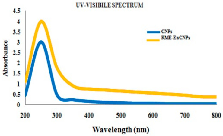
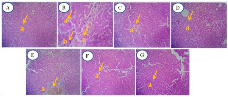
Alstonia venenata R.Br., a plant native to the Western Ghats, is recognized for its diverse medicinal properties. The plant's extracts, particularly rich in alkaloids and other bioactive compounds, have shown potential anticancer activity. This study investigates the therapeutic potential of chitosan nanoparticles (CNPs) loaded with the root methanolic extract (RME) of A. venenata in combating breast cancer induced by dimethylbenz(a)anthracene (DMBA) in female Sprague Dawley rats. The RME-loaded chitosan nanoparticles (RME-EnCNPs) were synthesized and characterized, and their in vivo efficacy was evaluated. Treatment with RME-EnCNPs significantly inhibited tumor progression, which is evidenced by reduced tumor volume, burden, and incidence. Moreover, the nanoparticles demonstrated a sustained release of the active compounds, leading to marked improvements in various biochemical, enzymatic, and histopathological parameters. The study found that both RME and RME-EnCNPs effectively suppressed tumor growth, with RME-EnCNPs showing superior efficacy in modulating tumor progression. Antioxidant assays revealed that treatment with RME-EnCNPs (500 mg/kg) resulted in significant increases in total protein, superoxide dismutase (SOD), catalase, glutathione peroxidase (GPx), and glutathione (GSH) levels, alongside a marked reduction in lipid peroxidation (LPO) (p < 0.001). These findings suggest that RME-EnCNPs exert a potent antioxidant effect, mitigating oxidative stress within the tumor microenvironment. The root extract of A. venenata and its nanoparticle formulation hold promise as a potential therapeutic agent for breast cancer, warranting further investigation to isolate active bioactive compounds and elucidate their mechanisms of action.

Keywords: Alstonia venenata; anticancer activity; antioxidant activity; chitosan nanoparticles; dimethylbenz(a)anthracene; rats.


Conflict of interest statement

The authors declare no conflicts of interest.

Figures

Figure 1
Figure 1
Entrapment efficiency of root methanolic extract-encapsulated chitosan nanoparticles (RME-EnCNPs). Effects a, b, c, d, and e were significant with consideration of probability p < 0.05. Values are means ± standard deviation of three replicates.
Figure 2
Figure 2
Drug release percentage of RME-EnCNPs.
Figure 3
Figure 3
In vitro drug release kinetic models.
Figure 4
Figure 4
UV absorption spectra of chitosan nanoparticles (CNPs) and root methanolic extract-encapsulated chitosan nanoparticles (RME-EnCNPs).
Figure 5
Figure 5
FTIR Spectra of chitosan nanoparticles (CNPs) and root methanolic extract-encapsulated chitosan nanoparticles (RME-EnCNPs).
Figure 6
Figure 6
X-ray diffraction pattern of chitosan nanoparticles (CNPs) at 2θ (degree).
Figure 7
Figure 7
X-ray diffraction pattern of root methanolic extract-encapsulated chitosan nanoparticles (RME-EnCNPs) at 2θ (degree).
Figure 8
Figure 8
SEM images of chitosan nanoparticles (CNPs) and root methanolic extract-encapsulated chitosan nanoparticles (RME-EnCNPs).
Figure 9
Figure 9
Zeta potential images of chitosan nanoparticles (CNPs) and root methanolic extract-encapsulated chitosan nanoparticles (RME-EnCNPs).
Figure 10
Figure 10
Effect of RME and RME-EnCNPs on hexokinase, aldolase, glucose-6-phosphatase, fructose 1-6-Di phosphatase levels in female Sprague Dawley rats. Results expressed by triplicate means (n = 3) ± standard error of mean (SEM). Statistical significance determined with Dunnett’s t-distribution with probability value, *** p < 0.001, ** p < 0.01, * p < 0.05 calculated by comparing treated group with control group. DMBA-7, 12-Dimethylbenz[a]anthracene; STD, standard (tamoxifen); RME, root methanolic extract; RME-EnCNPs, root methanolic extract-encapsulated chitosan nanoparticles.
Figure 11
Figure 11
Effect of root methanolic extract (RME) and root methanolic extract-encapsulated chitosan nanoparticles (RME-EnCNPs) on isocitrate dehydrogenase (IDH), succinate dehydrogenase (SDH), and malate dehydrogenase (MDH) levels in female Sprague Dawley rats. Results expressed by triplicate means (n = 3) ± standard error of mean (SEM). Statistical significance determined with Dunnett’s t-distribution with probability value, *** p < 0.001, ** p < 0.01, * p < 0.05 calculated by comparing treated group with control group. DMBA-7, 12-Dimethylbenz[a]anthracene; STD, standard (tamoxifen); RME, root methanolic extract; RME-EnCNPs, root methanolic extract-encapsulated chitosan nanoparticles.
Figure 12
Figure 12
Histology of DMBA-induced mammary tumors of female Sprague Dawley rats. (AG) Shows histopathological analysis of mammary tumors of control and experimental groups. Group (A): Control showed (a—arrow) normal architecture. Group (B): DMBA-induced cancer showed (a—arrow) malignant epithelial cells, (b—arrow) hyperchromasia, (c—arrow) nuclear cytoplasmic ratio and pleomorphism. Group (C): DMBA + standard (tamoxifen) treated animals showed (a—arrow) nipple areola complex, (b—arrow) hyperplastic glands and acinus, (c—arrow) areas of fibrosis and elastosis. Group (D): DMBA + RME 250 mg/kg treated animals showed (a—arrow) fibrosis with myxoid degeneration and individual cells of necrosis seen. Group (E): DMBA + RME 500 mg/kg showed (a—arrow) non-neoplastic glands and acinus, (b—arrow) fibrosis and occasional areas of necrosis seen. Group (F): VIDMBA + RME-EnCNPs 250 mg/kg treated animal shows (a—arrow) single layered non-neoplastic acinus. Group (G): DMBA + RME-EnCNPs 500 mg/kg treated animal shows (a—arrow) disappearance of tumor cells with no glands and acini noted, (b—arrow) fibrosis noted.
Figure 13
Figure 13
Histology of liver of DMBA-induced mammary tumor-bearing rats. (AG) Shows histopathological analysis of liver tissue of control and experimental groups. Group (A): control showed normal architecture of hepatocytes (a—arrow) normal architecture. Group (B): DMBA-induced cancer bearing animal showed (a—arrow) abnormal architecture, (b—arrow) hepatocytes arranged in cords, (c—arrow) portal tract appears remarkable. Group (C): DMBA + standard (tamoxifen) treated animals showed (a—arrow) central veins with congestion. Group (D): DMBA + RME 250 mg/kg showed (a—arrow) sinusoidal dilatation. Group (E): DMBA + RME 500 mg/kg showed (a—arrow) normal hepatocytes with sinusoidal dilatation. Group (F): DMBA + RME-EnCNPs 250 mg/kg treated animals showed (a—arrow) normal hepatocytes with portal tract. Group (G): DMBA + RME-EnCNPs 500 mg/kg treated animals showed (a—arrow) normal hepatocytes with central vein congestion.
Figure 14
Figure 14
Histology of kidney of DMBA-induced mammary tumor-bearing rats. (AG) Shows histopathological analysis of kidney tissue of control and experimental groups. Group (A): control showed (a—arrow) normal architecture. Group (B): DMBA-induced cancer bearing animals showed (a—arrow) glomeruli, (b—arrow) loss of mesangial matrix expansion. Group (C): DMBA + standard (tamoxifen) treated animals showed (a—arrow) normal glomeruli. Group (D): DMBA + RME 250 mg/kg showed (a—arrow) focal mild eosinophilic material in lumen. Group (E): DMBA + RME 500 mg/kg showed (a—arrow) no significant pathology in glomeruli. Interstitium showed normal. VI Group (F): DMBA + RME-EnCNPs 250 mg/kg treated animals showed (a—arrow) blood vessels with mild congestion. Group (G): DMBA + RME-EnCNPs 500 mg/kg treated animals showed (a—arrow) normal morphology in interstitium.
Figure 15
Figure 15
Histology of uterus of DMBA-induced mammary tumor-bearing rats. (AG) Shows histopathological analysis of uterus tissue of control and experimental groups. Group (A): control showed (a—arrow) normal architecture. Group (B): DMBA-induced cancer bearing animals showed (a—arrow) abnormal epithelium with scattered inflammatory infiltrates. Group (C): DMBA + standard (tamoxifen) treated animals showed (a—arrow) glands with circular and elongated shape. Group (D): DMBA + RME 250 mg/kg showed (a—arrow) stroma with scattered inflammatory infiltrates. Group (E): DMBA + RME 500 mg/kg showed (a—arrow) normal mucosa. Group (F): DMBA + RME-EnCNPs 250 mg/kg treated animals showed (a—arrow) neutrophils and eosinophils. Group (G): DMBA + RME-EnCNPs 500 mg/kg treated animals showed (a—arrow) normal endomyometrium with scattered infiltrates.

Similar articles

References

    1. Ferlay J., Colombet M., Soerjomataram I., Parkin D.M., Piñeros M., Znaor A., Bray F., Global Cancer Observatory: Cancer Today [Internet] Lyon: International Agency for Research on Cancer. 2018. [(accessed on 1 June 2022)]. Available online: https://gco.iarc.fr/today/
    1. Arnold M., Morgan E., Rumgay H., Mafra A., Singh D., Laversanne M., Vignat J., Gralow J.R., Cardoso F., Siesling S., et al. Current and future burden of breast cancer: Global statistics for 2020 and 2040. Breast. 2022;66:15–23. doi: 10.1016/j.breast.2022.08.010. - DOI - PMC - PubMed
    1. Gullino P.M., Pettigrew H.M., Grantham F.H. N-nitrosomethylurea as mammary gland carcinogen in rats. J. Natl. Cancer Inst. 1975;54:401–414. doi: 10.1093/jnci/54.2.401. - DOI - PubMed
    1. Anderson M.E., Meister A. Transport and direct utilization of gamma-glutamylcyst (e) ine for glutathione synthesis. Proc. Natl. Acad. Sci. USA. 1983;80:707–711. doi: 10.1073/pnas.80.3.707. - DOI - PMC - PubMed
    1. Mottaghitalab F., Farokhi M., Fatahi Y., Atyabi F., Dinarvand R. New insights into designing hybrid nanoparticles for lung cancer: Diagnosis and treatment. J. Control. Release. 2019;295:250–267. doi: 10.1016/j.jconrel.2019.01.009. - DOI - PubMed

LinkOut - more resources