. 2024 Dec 19;13(12):1562.
doi: 10.3390/antiox13121562.

Antioxidant, Anti-Inflammatory, and Anticancer Activities of Five Citrus Peel Essential Oils

Affiliations

Antioxidant, Anti-Inflammatory, and Anticancer Activities of Five Citrus Peel Essential Oils

Yurong Li et al. Antioxidants (Basel). .

Abstract

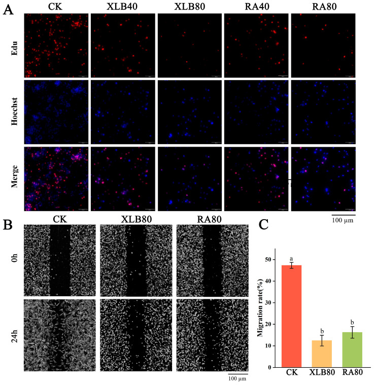
Citrus peel essential oil (CPEO) is favored by people for its aromatic scent, while also possessing numerous bioactive compounds that are advantageous to human health. This study evaluated the antioxidant, anti-inflammatory, and anticancer activities of CPEOs through cell experiments. The results showed that CPEOs could increase the activity of the antioxidant enzyme system and nonenzymatic defence system in H2O2-treated RAW 264.7 cells by reducing cellular lipid peroxidation. CPEOs also reduced the nitric oxide production induced by lipopolysaccharide treatment in RAW 264.7 cells while decreasing proinflammatory cytokines expression and increasing anti-inflammatory cytokine expression. Wound healing assays, flow cytometry, and quantitative real-time fluorescent quantitative PCR (qRT-PCR) revealed that CPEOs could induce apoptosis in U87 cells through the mitochondrial apoptotic pathway. These findings indicate that CPEOs possess excellent antioxidant, anti-inflammatory, and anticancer activity potential, making them suitable for use in functional antioxidant and anti-inflammatory foods and nutritional health products.

Keywords: anti-inflammatory; anticancer; antioxidants; citrus; essential oil.


Conflict of interest statement

The authors declare that they have no known competing financial interests or personal relationships that could have appeared to influence the work reported in this paper.

Figures

Figure 1
Figure 1
The effect of five CPEOs on the cell viability of RAW264.7 cells and U87 cells. (A) The changes in cell viability after treating RAW264.7 cells with different concentrations of FJ, JC26, STJ, XLB, and RA EOs for 24 h. (B) Treatment with 0.5% DMSO did not affect the viability of RAW264.7 cells. (CG) The changes in cell viability after treating U87 cells with different concentrations of FJ, JC26, STJ, XLB, and RA EOs for 24 h. (H) Treatment with 0.5% DMSO did not affect the viability of U87 cells. The data represent the mean ± SD. The blue dashed line indicated a cell viability rate of 50%.
Figure 2
Figure 2
T The effect of CPEOs on cell viability (A), MDA (B), GSH (C), GR (D), CAT (E), and SOD (F) in RAW264.7 cells after induction with 0.03% H2O2 for 2 h. CK was treated with 0.5% DMSO. The concentration of AsA in the positive control was 25 µg/mL. The concentration of the five CPEOs was 50 μg/mL. The data represent the mean ± SD. “pro” represents protein. Values followed by different superscripts (a–g) are significantly different (p < 0.05).
Figure 3
Figure 3
The effect of CPEOs on the production of NO (A), IL-6 (B), TNF-α (C), IFN-γ (D), and IL-10 (E) in RAW264.7 cells after induction with 3 μg/mL LPS for 4 h. CK was treated with 0.5% DMSO. The concentration of DEX in the positive control was 5 µg/mL. The concentration of the five CPEOs was 50 μg/mL. The data represent the mean ± SD. Values followed by different superscripts (a–g) are significantly different (p < 0.05).
Figure 4
Figure 4
Effect of the CPEOs on proliferation and migration in U87 cells. (A) XLB and RA EOs can inhibit cell proliferation. Proliferating cells and cell nuclei were stained with BeyoClick™ EdU-555 kit and Hoechst 33342. (B) The cell migration ability of U87 cells was determined using the wound healing assay, where the cells were treated with XLB and RA EOs in serum-free medium for 24 h. (C) Statistical data of migration rate (%). The concentrations of XLB40 and RA40 were 40 µg/mL, while those of XLB80 and RA80 were 80 µg/mL. CK was treated with 0.5% DMSO. The data represent the mean ± SD. Values followed by different superscripts (a,b) are significantly different (p < 0.05).
Figure 5
Figure 5
Effects of the CPEOs on the U87 cells cycle. (AE) Determination of cell cycle distribution by flow cytometry. (F) The proportions of cell populations in G0/G1, S, and G2/M phases after treatment with XLB and RA EOs. The concentrations of XLB40 and RA40 were 40 µg/mL, while those of XLB80 and RA80 were 80 µg/mL. CK was treated with 0.5% DMSO. The data are presented as mean ± SD. Values followed by different superscripts (a–e) are significantly different (p < 0.05).
Figure 6
Figure 6
Effects of the CPEOs on U87 cell apoptosis. (AE) Apoptotic cells were assessed by flow cytometry after Annexin V-FITC and PI staining. Q1 represents necrotic cells; Q2 represents late apoptotic cells; Q3 represents early apoptotic cells; Q4 represents viable cells. (F) Living cells, early and late apoptotic cell rates. The concentrations of XLB40 and RA40 were 40 µg/mL, while those of XLB80 and RA80 were 80 µg/mL. CK was treated with 0.5% DMSO. The data are presented as mean ± SD. Values followed by different superscripts (a–e) are significantly different (p < 0.05).
Figure 7
Figure 7
Effect of the CPEOs on the MMP and the relative expression levels of apoptosis-related genes in U87 cells. (A) Fluorescence images of U87 incubated with JC-1 after treatment with XLB and RA EOs. (B) MMP quantified by measuring green fluorescence intensity and red fluorescence intensity. (C) BAX; (D) Caspase-9; (E) Caspase-7; (F) Caspase-3. The concentrations of XLB40 and RA40 were 40 µg/mL, while those of XLB80 and RA80 were 80 µg/mL. CK was treated with 0.5% DMSO. The data are presented as mean ± SD. Values followed by different superscripts (a–e) are significantly different (p < 0.05).

Similar articles

References

    1. Suri S., Singh A., Nema P.K. Current Applications of Citrus Fruit Processing Waste: A Scientific Outlook. Appl. Food Res. 2022;2:100050. doi: 10.1016/j.afres.2022.100050. - DOI
    1. Ben Hsouna A., Sadaka C., Generalić Mekinić I., Garzoli S., Švarc-Gajić J., Rodrigues F., Morais S., Moreira M.M., Ferreira E., Spigno G., et al. The Chemical Variability, Nutraceutical Value, and Food-Industry and Cosmetic Applications of Citrus Plants: A Critical Review. Antioxidants. 2023;12:481. doi: 10.3390/antiox12020481. - DOI - PMC - PubMed
    1. Ambrosio C.M.S., Diaz-Arenas G.L., Agudelo L.P.A., Stashenko E., Contreras-Castillo C.J., da Gloria E.M. Chemical Composition and Antibacterial and Antioxidant Activity of a Citrus Essential Oil and Its Fractions. Molecules. 2021;26:2888. doi: 10.3390/molecules26102888. - DOI - PMC - PubMed
    1. Palma C.E., Cruz P.S., Cruz D.T.C., Bugayong A.M.S., Castillo A.L. Chemical Composition and Cytotoxicity of Philippine Calamansi Essential Oil. Ind. Crop. Prod. 2019;128:108–114. doi: 10.1016/j.indcrop.2018.11.010. - DOI
    1. Singh B., Singh J.P., Kaur A., Yadav M.P. Insights into the Chemical Composition and Bioactivities of Citrus Peel Essential Oils. Food Res. Int. 2021;143:110231. doi: 10.1016/j.foodres.2021.110231. - DOI - PubMed

LinkOut - more resources