. 2024 Dec 23;13(12):1585.
doi: 10.3390/antiox13121585.

Neuroprotective Potential of Indole-Based Compounds: A Biochemical Study on Antioxidant Properties and Amyloid Disaggregation in Neuroblastoma Cells

Affiliations

Neuroprotective Potential of Indole-Based Compounds: A Biochemical Study on Antioxidant Properties and Amyloid Disaggregation in Neuroblastoma Cells

Tania Ciaglia et al. Antioxidants (Basel). .

Abstract

Based on the established neuroprotective properties of indole-based compounds and their significant potential as multi-targeted therapeutic agents, a series of synthetic indole-phenolic compounds was evaluated as multifunctional neuroprotectors. Each compound demonstrated metal-chelating properties, particularly in sequestering copper ions, with quantitative analysis revealing approximately 40% chelating activity across all the compounds. In cellular models, these hybrid compounds exhibited strong antioxidant and cytoprotective effects, countering reactive oxygen species (ROS) generated by the Aβ(25-35) peptide and its oxidative byproduct, hydrogen peroxide, as demonstrated by quantitative analysis showing on average a 25% increase in cell viability and a reduction in ROS levels to basal states. Further analysis using thioflavin T fluorescence assays, circular dichroism, and computational studies indicated that the synthesized derivatives effectively promoted the self-disaggregation of the Aβ(25-35) fragment. Taken together, these findings suggest a unique profile of neuroprotective actions for indole-phenolic derivatives, combining chelating, antioxidant, and anti-aggregation properties, which position them as promising compounds for the development of multifunctional agents in Alzheimer's disease therapy. The methods used provide reliable in vitro data, although further in vivo validation and assessment of blood-brain barrier penetration are needed to confirm therapeutic efficacy and safety.

Keywords: amyloid; antioxidants; disaggregation; in-cell studies; indole nucleus; neuroprotection.


Conflict of interest statement

The authors declare no conflicts of interest.

Figures

Figure 1
Figure 1
Scheme of the synthesized molecules.
Figure 2
Figure 2
(A) UV spectra (in the range of 280 to 400 nm) of compounds (30 µM) alone and in the presence of 40 µM FeSO4, FeCl3, and CuSO4. (B) Copper-chelating quantitative analyses of compounds (30 µM). EDTA (1 mM) was used as the positive control. Results are shown as mean ± standard deviation (SD) from three independent experiments. **, *** denote p < 0.01 and p < 0.001 vs. Ctrl.
Figure 3
Figure 3
Neuroprotective activity of compounds. (A) SH-SY5Y cells were exposed to compounds at a concentration of 30 µM. Neuroprotective effects of compounds against (B) H2O2-induced (500 μM) cytotoxicity, (C) H2O2-induced (500 μM) ROS production, and (D) Aβ(25–35)-induced (40 μM) cytotoxicity. The 2′,7′-dichlorofluorescin diacetate (DCFH-DA) assay was conducted to reveal ROS production. The changes in viability were determined by calculating the percentage of viable cells in treated cultures relative to untreated controls. The results are presented as the mean ± standard deviation (SD) from three independent experiments. *, **, and *** denote, respectively, p < 0.05, p < 0.01, and p < 0.001 vs. Ctrl; ##, and ### denote, respectively, p < 0.01, and p < 0.001 vs. H2O2 or Aβ(25–35).
Figure 4
Figure 4
Disaggregating properties of compounds. In-cell ThT assay was performed for both fluorescence microscopy (A) and spectrophotometry (B). Scale bar: 100 μm. (N ≥ 10). Cells were observed at 20× magnification. (C) ThT shows a direct disaggregating effect of compounds against Aβ. Data are shown as mean ± SD of three different experiments performed in triplicate. *** denotes p < 0.001 vs. Ctrl; ##, and ### denote, respectively, p < 0.01, and p < 0.001 vs. H2O2 or Aβ(25–35).
Figure 5
Figure 5
(A,B) CD curves and secondary structure analysis of the Aβ(25−35) peptide were performed using the CONTIN algorithm after 24 h of aggregation. Aβ(25–35) 40 μM was used as a positive control. A different colour has been selected for Aβ(25–35) and each compound in presence Aβ(25–35), as indicated in the legend.
Figure 6
Figure 6
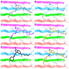
Three-dimensional model of the interactions of 12 (A), 13 (B), 14 (C), 20 (D), 21 (E), and 22 (F) with Aβ(25–35). The biological target is depicted by a ribbon colored by a chain (D, magenta; E, azure; F, green; G, faded red) and tube (colored: C, by chain; polar H, white; N, dark blue; O, red; S, yellow). The small molecules are represented by sticks (gray for 12, cyan for 13, violet for 14, orange for 20, black for 21, khaki for 22) and balls (colored: C, as for the sticks; polar H, white; N, dark blue; O, red). The hydrogen bonds between the ligand and Aβ(25–35) are represented by the dashed black lines.

Similar articles

Cited by

References

    1. Sies H. Strategies of Antioxidant Defense. Eur. J. Biochem. 1993;215:213–219. doi: 10.1111/j.1432-1033.1993.tb18025.x. - DOI - PubMed
    1. Di Meo S., Reed T.T., Venditti P., Victor V.M. Role of ROS and RNS Sources in Physiological and Pathological Conditions. Oxid. Med. Cell. Longev. 2016;2016:1245049. doi: 10.1155/2016/1245049. - DOI - PMC - PubMed
    1. Liguori I., Russo G., Curcio F., Bulli G., Aran L., Della-Morte D., Gargiulo G., Testa G., Cacciatore F., Bonaduce D. Oxidative Stress, Aging, and Diseases. Clin. Interv. Aging. 2018;13:757–772. doi: 10.2147/CIA.S158513. - DOI - PMC - PubMed
    1. Rahimi H., Mirnezami M., Yazdabadi A. Bilirubin as a New Antioxidant in Melasma. J. Cosmet. Dermatol. 2022;21:5800–5803. doi: 10.1111/jocd.15240. - DOI - PubMed
    1. Cruz-Sánchez F.F., Gironès X., Ortega A., Alameda F., Lafuente J.V. Oxidative Stress in Alzheimer’s Disease Hippocampus: A Topographical Study. J. Neurol. Sci. 2010;299:163–167. doi: 10.1016/j.jns.2010.08.029. - DOI - PubMed

Grants and funding

LinkOut - more resources