. 2024 Dec 16;16(24):4194.
doi: 10.3390/cancers16244194.

Chemokine CXCL12 Activates CXC Receptor 4 Metastasis Signaling Through the Upregulation of a CXCL12/CXCR4/MDMX (MDM4) Axis

Affiliations

Chemokine CXCL12 Activates CXC Receptor 4 Metastasis Signaling Through the Upregulation of a CXCL12/CXCR4/MDMX (MDM4) Axis

Rusia Lee et al. Cancers (Basel). .

Abstract

Background: The metastasis-promoting G-protein-coupled receptor CXC Receptor 4 (CXCR4) is activated by the chemokine CXCL12, also known as stromal cell-derived factor 1 (SDF-1). The CXCL12/CXCR4 pathway in cancer promotes metastasis but the molecular details of how this pathway cross-talks with oncogenes are understudied. An oncogene pathway known to promote breast cancer metastasis in MDA-MB-231 xenografts is that of Mouse Double Minute 2 and 4 (MDM2 and MDM4, also known as MDMX). MDM2 and MDMX promote circulating tumor cell (CTC) formation and metastasis, and positively correlate with a high expression of CXCR4. Interestingly, this MDMX-associated upregulation of CXCR4 is only observed in cells grown in the tumor microenvironment (TME), but not in MDA-MB-231 cells grown in a tissue culture dish. This suggested a cross-talk signaling factor from the TME which was predicted to be CXCL12 and, as such, we asked if the exogenous addition of the cell non-autonomous CXCL12 ligand would recapitulate the MDMX-dependent upregulation of CXCR4.

Methods: We used MDA-MB-231 cells and isolated CTCs, with and without MDMX knockdown, plus the exogenous addition of CXCL12 to determine if MDMX-dependent upregulation of CXCR4 could be recapitulated outside of the TME context. We added exogenous CXCL12 to the culture medium used for growth of MDA-MB-231 cells and isogenic cell lines engineered for MDM2 or MDMX depletion. We carried out immunoblotting, and quantitative RT-PCR to compare the expression of CXCR4, MDM2, MDMX, and AKT activation. We carried out Boyden chamber and wound healing assays to assess the influence of MDMX and CXCL12 on the cells' migration capacity.

Results: The addition of the CXCL12 chemokine to the medium increased the CXCR4 cellular protein level and activated the PI3K/AKT signaling pathway. Surprisingly, we observed that the addition of CXCL12 mediated the upregulation of MDM2 and MDMX at the protein, but not at the mRNA, level. A reduction in MDMX, but not MDM2, diminished both the CXCL12-mediated CXCR4 and MDM2 upregulation. Moreover, a reduction in both MDM2 and MDMX hindered the ability of the added CXCL12 to promote Boyden chamber-assessed cell migration. The upregulation of MDMX by CXCL12 was mediated, at least in part, by a step upstream of the proteasome pathway because CXCL12 did not increase protein stability after cycloheximide treatment, or when the proteasome pathway was blocked.

Conclusions: These data demonstrate a positive feed-forward activation loop between the CXCL12/CXCR4 pathway and the MDM2/MDMX pathway. As such, MDMX expression in tumor cells may be upregulated in the primary tumor microenvironment by CXCL12 expression. Furthermore, CXCL12/CXCR4 metastatic signaling may be upregulated by the MDM2/MDMX axis. Our findings highlight a novel positive regulatory loop between CXCL12/CXCR4 signaling and MDMX to promote metastasis.

Keywords: CXCR4; MDM2; MDMX; PI3K/AKT signaling; chemokine signaling; circulating tumor cells (CTCs); metastasis; triple-negative breast cancer (TNBC); tumor microenvironment (TME).


Conflict of interest statement

The authors declare no conflicts of interest.

Figures

Figure 1
Figure 1
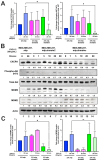
Chemokine CXCL12 addition to cell culture increases CXCR4, activates PI3K/AKT, and MDMX and MDM2 protein levels. (A) Panels show immunoblot analysis for MDMX, MDM2, CXCR4, and phospho-AKT S473, total AKT, and actin in MDA-MB-231.mlp cells after treatment for up to 60 min with CXCL12 at 50 ng/mL for 0, 1, 5, 10, 20, and 60 min, lanes 1–6. Total AKT and actin were used as loading controls. The proteins were derived from the same samples run on different gels/membranes at the same time and their molecular weights are shown. (B) Untreated cells and those following the 20 min treatments were compared for the MDMX and MDM2 protein levels evaluated (using actin as a normalizer control for loading) using ImageJ and graphs were created with Prism 10 software, with the untreated value set as 1 and the ratio reported for CXCL12-treated samples (20 min) used to report the fold change. (C) Untreated and 20 min treatments were compared for CXCR4 and phospho-AKT S473 protein levels quantified via ImageJ relative to total AKT as a loading control with the untreated value set as 1 and the ratio reported for CXCL12-treated samples (20 min) used to report the fold change. Images were analyzed using ImageJ and graphs were created with Prism software. Error bars represent SD. * p < 0.05, ** p < 0.01, NS = non-significant (N = 4 biological replicates).
Figure 2
Figure 2
Knockdown of MDMX in MDA-MB-231 cells disrupts CXCL12 signaling to upregulate CXCR4, MDM2, and AKT activation. MDA-MB-231.mlp, MDA-MB-231.mlp.shmdm2, and MDA-MB-231.mlp.shmdmx cells treated in cell culture with the addition of CXCL12 at a final concentration of 50 ng/mL in cell culture for up to 60 min. (A) CXCR4 and phospho-AKT S473 protein levels were semi-quantified at 20 min via ImageJ relative to actin as a loading control. Protein level analysis was carried out using Western blot results using Image J and Prism software and densitometries were measured as a ratio relative to the actin band density. Fold change was calculated relative to protein levels in the untreated 231.mlp vector control cells. Error bars represent SD. * p < 0.05, NS = non-significant (N = 3 biological replicates). (B) Immunoblot analysis for CXCR4, phospho-AKT, MDMX, and MDM2 protein levels in MDA-MB-231 or knockdown cells after the addition of CXCL12. (C) MDMX and MDM2 protein levels were semi-quantified at 20 min post addition of CXCL12 to the cell culture. Protein levels were normalized to actin and fold change was calculated relative to untreated 231.mlp vector control cells. MDM2 or MDMX knockdown were confirmed for each respective cell line. Protein level analysis was carried out from Western blot results using Image J and Prism software and expression scores were normalized to actin. Error bars represent SD. * p < 0.05, NS = non-significant (N = 3 biological replicates).
Figure 3
Figure 3
CXCL12 does not enhance chemotaxis in MDM2- or MDMX-knockdown MDA-MB-231 cells. (A) Representative images of crystal violet-stained cells of chemotaxis assay membrane inserts. A total of 50,000 MDA-MB-231 cells were loaded into the upper chamber in media (final vol: 200 µL). Migration was initiated by adding 500 µL of medium to the lower chamber with or without CXCL12 at a final concentration of 50 ng/mL for 24 h. Cells were incubated for 24 h at 37 degree Celsius in a 5% CO2 incubator. Insert was stained with crystal violet and washed with Millipore water. Representative images of stained cells on insert shown by imaging via microscopy. (B) Graph of % wound closure at the 24 h time point. Error bars represent SD. * p < 0.05, *** p < 0.001, NS = nonsignificant. The p values were calculated using two-tailed unpaired t tests on Prism software.
Figure 4
Figure 4
MDA-MB-231.mlp.CTC lines compared to MDA-MB-231.shmdm2.CTC and MDA-MB-231.shmdmx.CTC maintain increased migratory compacity but have reduced response to CXCL12. (A) Immunoblot of whole cell lysates from MDA-MB-231.mlp, MDA-MB-231.shmdm2, and MDA-MB-231.shmdmx (lanes 1–3) and MDA-MB-231.mlp.CTC A and B (lanes 4 and 5), MDA-MB-231.shmdm2.CTC (lane 6), and MDA-MB-231.shmdmx.CTC (lane 7) cell lines probed for MDMX (top) and MDM2 (middle). The loading control was actin (bottom) (B) Representative images of MDA-MB-231-derived CTC lines in wound healing assay. Black lines denote the borders of the scratch made. (C) Graph of % wound closure at 12 h time point. Error bars represent SD. *** p < 0.001, NS = nonsignificant. The p value was calculated using two-tailed unpaired t tests on Prism software (D) Immunoblot of whole cell lysates from MDA-MB-231.mlp.CTC lines treated with CXCL12 at a final concentration of 50 ng/mL for 30 and 60 min. (E) MDMX and MDM2 protein expression was compared using ImageJ quantitation relative to actin, respectively, as a loading control.
Figure 5
Figure 5
CXCL12 addition does not increase MDMX protein half-life following cycloheximide or MG132 treatment. (A) Immunoblot analysis of lysates from MDA-MB-231 cells treated with CXCL12 (50 ng/mL) for 30 min followed by cycloheximide (CHX) or DMSO for 40 or 80 min. Cells were harvested and lysed in CHAPS lysis buffer and subjected to immunoblotting to probe for MDM2 and MDMX. Actin was probed as a loading control. (B,C) Evaluation of Western blot band density was carried out using ImageJ and Prism software. Error bars represent SD. * p < 0.05, NS = non-significant. (D) HCT116 p53-/- cells were transfected with pcDNA3-MDMX for 24 h and then treated with CXCL12 (50 ng/mL) for 30 min followed by MG132 or DMSO for 40 or 80 min. Cells were harvested and lysed in CHAPS lysis buffer and subjected to immunoblotting to probe for MDM2, MDMX, and Ubiquitin. Actin was probed as a loading control. (E,F) Evaluation of Western blot band density was carried out using ImageJ and Prism 10 software. Error bars represent SD. * p < 0.05, NS = non-significant.
Figure 6
Figure 6
A model of CXCL12/CXCR4/AKT signaling to the MDMX/MDM2 axis. CXCL12/CXCR4/AKT signaling works to upregulate MDMX in the primary tumor in cooperation with the TME and results in a feed forward activation loop. This promotes intravasation of the cancer cells into the blood stream and the promotion of CTCs. The CTCs are then able to survive but have a reduced CXCL12 feed-forward loop with MDMX and MDM2 which we posit is re-established when cells extravasate into CXCL12-rich environments to form metastasis.

References

    1. Hauck P.M., Wolf E.R., Olivos D.J., 3rd, Batuello C.N., McElyea K.C., McAtarsney C.P., Cournoyer R.M., Sandusky G.E., Mayo L.D. Early-Stage Metastasis Requires Mdm2 and Not p53 Gain of Function. Mol. Cancer Res. 2017;15:1598–1607. doi: 10.1158/1541-7786.MCR-17-0174. - DOI - PMC - PubMed
    1. Muller A., Homey B., Soto H., Nianfeng G., Catron D., Buchanan M.E., McClanahan T., Murphy E., Yuan W., Wagner S.N., et al. Involvement of chemokine receptors in breast cancer metastasis. Nature. 2001;410:50–56. doi: 10.1038/35065016. - DOI - PubMed
    1. Gao C., Xiao G., Bargonetti J. Contemplations on MDMX (MDM4) driving triple negative breast cancer circulating tumor cells and metastasis. Oncotarget. 2019;10:5007–5010. doi: 10.18632/oncotarget.27134. - DOI - PMC - PubMed
    1. Gao C., Xiao G., Piersigilli A., Gou J., Ogunwobi O., Bargonetti J. Context-dependent roles of MDMX (MDM4) and MDM2 in breast cancer proliferation and circulating tumor cells. Breast Cancer Res. 2019;21:5. doi: 10.1186/s13058-018-1094-8. - DOI - PMC - PubMed
    1. Xin Y., Hu B., Li K., Hu G., Zhang C., Chen X., Tang K., Du P., Tan Y. Circulating tumor cells with metastasis-initiating competence survive fluid shear stress during hematogenous dissemination through CXCR4-PI3K/AKT signaling. Cancer Lett. 2024;590:216870. doi: 10.1016/j.canlet.2024.216870. - DOI - PubMed

LinkOut - more resources