Review
. 2024 Dec 18;16(24):4212.
doi: 10.3390/cancers16244212.

The Functional Interaction Between Epstein-Barr Virus and MYC in the Pathogenesis of Burkitt Lymphoma

Affiliations
Review

The Functional Interaction Between Epstein-Barr Virus and MYC in the Pathogenesis of Burkitt Lymphoma

Sandra Solares et al. Cancers (Basel). .

Abstract

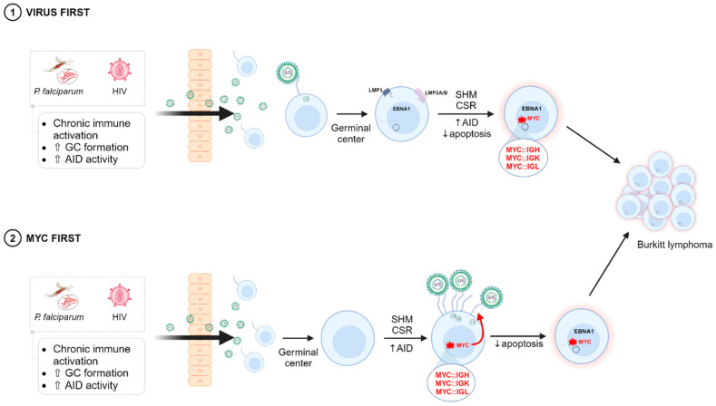
The Epstein-Barr virus (EBV) is associated with a wide range of diseases, malignant and non-malignant. EBV was, in fact, the first virus described with cell transformation capacity, discovered by Epstein in 1964 in lymphoma samples from African children. Since then, EBV has been associated with several human tumors including nasopharyngeal carcinoma, gastric carcinoma, T-cell lymphoma, Hodgkin lymphoma, diffuse large B cell lymphoma, and Burkitt lymphoma among others. The molecular hallmark of Burkitt lymphoma (BL) is a chromosomal translocation that involves the MYC gene and immunoglobulin loci, resulting in the deregulated expression of MYC, an oncogenic transcription factor that appears deregulated in about half of human tumors. The role of MYC in lymphoma is well established, as MYC overexpression drives B cell proliferation through multiple mechanisms, foremost, the stimulation of the cell cycle. Indeed, MYC is found overexpressed or deregulated in several non-Hodgkin lymphomas. Most endemic and many sporadic BLs are associated with EBV infection. While some mechanisms by which EBV can contribute to BL have been reported, the mechanism that links MYC translocation and EBV infection in BL is still under debate. Here, we review the main EBV-associated diseases, with a special focus on BL, and we discuss the interaction of EBV and MYC translocation during B cell malignant transformation in BL.

Keywords: Burkitt lymphoma; Epstein–Barr virus; MYC.


Conflict of interest statement

The authors declare no conflicts of interest.

Figures

Figure 1
Figure 1
Epstein–Barr virus (EBV) structure and genome. (a) Scheme depicting the most relevant features of the EBV structure. Ø: virion diameter. (b) Schematic representation of the EBV linearized genome. Green and red boxes represent the EBNAs and LMPs latent genes, respectively. Violet arrows represent ncRNAs. The four alternative promoters from which EBNA genes are transcribed are represented by colored arrows (Cp, Wp, Fp, Qp). OriP: Replication origin. TR: Terminal Repeats. Created in BioRender. Garcia, L. (2024) BioRender.com/y04j261.
Figure 2
Figure 2
EBV latency programs and associated malignancies. After primary infection, the infected cell enters the Latency III program, also known as the growth program, expressing all latent genes, BARTs, and EBERs. This activates B cells promoting their transition toward a germinal center reaction and switching to Latency II or the default program, with a more restricted gene expression pattern. Finally, memory B cells exit the germinal center showing the Latency 0 program, also known as “Persistence”, in which only ncRNAs are expressed. Eventually, the transition from Latency 0 to Latency I (or the EBNA-1 only program) may occur when memory B cells proliferate so that the EBV episome is properly segregated to the daughter cells. In blue, EBV-associated lymphomas with their corresponding latency program. In red, EBV-associated epithelial malignancies and their corresponding latency programs are included. Created in BioRender. Garcia, L. (2024) BioRender.com/y04j261.
Figure 3
Figure 3
Molecules implicated in the interaction and entry of EBV with its host cell. (a) Scheme depicting EBV interaction with B cells. gp350/220 from the EBV envelope interacts with CR2 (or CR1), facilitating gp42 interaction with the HLA class II of the B cell surface. This triggers EBV fusion with the B cell membrane through gB to further deliver the virus genoma inside the cell. PDB: 8SM0 (structure of gp350/220 in complex with CR2). PDB: 1KG0 (Epstein–Barr virus gp42 bound to the MHC class II Receptor HLA-DR1). (b) Scheme depicting the EBV interaction with epithelial cells. gp350/220 or BMRF2 recognizing CR2 or β1-integrins, respectively. gH/gL interact with different β5, β6, and β8 integrins and MYH9 in the cell surface. gB, which drives the fusion with the cell membrane, interacts with NRP1. EPH2A binds both gH/gL and gB. Created in BioRender. Garcia, L. (2024) BioRender.com/y04j261.
Figure 4
Figure 4
The oncogenic MYC transcription factor and its translocations within the different Ig loci found in Burkitt lymphoma. (a) Scheme of the MYC protein. The conserved boxes among MYC proteins are in green. The bHLHLZ domain is in red. The most prevalent mutations in BL are indicated. TAD, transactivation domain; PEST, degradation domain; NLS, nuclear localization signals. (b). Schematic representation (not to scale) of the breakpoints in the chromosome 8 in BL. The three exons of the MYC genes are the blue boxes, with the coding regions in darker blue. The major promoter, P2, is shown. The three classes of breakpoints with the chromosome 14 (immunoglobulin heavy chain genes) are indicated as class I, II, and III and the BL subtype where it is more prevalent. The breakpoint in the eBL typically occurs > 10 kb upstream of the first MYC exon and in the first noncoding exon or first intron in the sBL. (c) Schematic figures (not to scale) of prototypical translocations MYC::IGH to heavy chain genes (left schemes, 80% of the BL cases), MYC::IGK to light chain kappa genes (upper right scheme, 15%), and MYC::IGL light chain lambda genes (lower right scheme, 5%). In the t(8;14) translocation, the break points are the switch regions of the constant gene segments encoding the IgH isotypes and are upstream of MYC. In eBL, the breakpoint in the chromosome 14 is more frequently in the J segment. The predominant breakpoint in sBL occurs in the Cµ (M) regions of the H chain locus. In the t(2;8) and t(8;22) translocations, the kappa and lambda light chain lie far downstream of MYC. AP refers to an alternative promoter used in this type of translocations. The enhancers are denoted as red circles. Adapted from [163,170]. Partially Created in BioRender. Garcia, L. (2024) BioRender.com/y04j261.
Figure 5
Figure 5
Two alternative models of EBV infection and MYC translocation in B cells prior to Burkitt lymphoma development. Chronic immune activation triggered by malaria or HIV leads to increased B cell proliferation, germinal center formation, and AID activity. In the virus first model, higher loads of EBV due to the weakened immune system favor EBV-mediated infection of B cells, leading to the increased susceptibility of MYC translocations during somatic hypermutation (SHM) and class switch recombination (CSR) in the germinal center. In the MYC first model, the chronic immune activation due to malaria or HIV makes the B cells prone to MYC translocations due to errors during SHM or CSR. Translocated MYC would lead to higher CR2 density on the surface of B cells, increasing the probability of EBV infection. In both models, EBV would confer the resistance mechanisms to apoptosis needed for the B cell to cope with the deregulated MYC activity, leading to Burkitt lymphoma development. Created in BioRender. Garcia, L. (2024) BioRender.com/y04j261.

Similar articles

Cited by

References

    1. Epstein M.A., Achong B.G., Barr Y.M. Virus particles in cultured lymphoblasts from Burkitt’s lymphoma. Lancet. 1964;1:702–703. doi: 10.1016/S0140-6736(64)91524-7. - DOI - PubMed
    1. Sample J., Young L., Martin B., Chatman T., Kieff E., Rickinson A., Kieff E. Epstein-Barr virus types 1 and 2 differ in their EBNA-3A, EBNA-3B, and EBNA-3C genes. J. Virol. 1990;64:4084–4092. doi: 10.1128/jvi.64.9.4084-4092.1990. - DOI - PMC - PubMed
    1. Gratama J.W., Ernberg I. Molecular epidemiology of Epstein-Barr virus infection. Adv. Cancer Res. 1995;67:197–255. - PubMed
    1. Zimber U., Adldinger H.K., Lenoir G.M., Vuillaume M., Knebel-Doeberitz M.V., Laux G., Desgranges C., Wittmann P., Freese U.K., Schneider U., et al. Geographical prevalence of two types of Epstein-Barr virus. Virology. 1986;154:56–66. doi: 10.1016/0042-6822(86)90429-0. - DOI - PubMed
    1. de-Thé G., Day N.E., Geser A., Lavoué M.F., Ho J.H., Simons M.J., Sohier R., Tukei P., Vonka V., Zavadova H. Sero-epidemiology of the Epstein-Barr virus: Preliminary analysis of an international study—A review. IARC Sci. Publ. 1975;11:3–16. - PubMed

LinkOut - more resources