. 2024 Nov 21;14(12):1482.
doi: 10.3390/biom14121482.

Thyroid Carcinoma Glycoproteins Express Altered N-Glycans with 3-O-Sulfated Galactose Residues

Affiliations

Thyroid Carcinoma Glycoproteins Express Altered N-Glycans with 3-O-Sulfated Galactose Residues

Jordan M Broekhuis et al. Biomolecules. .

Abstract

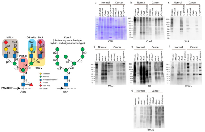
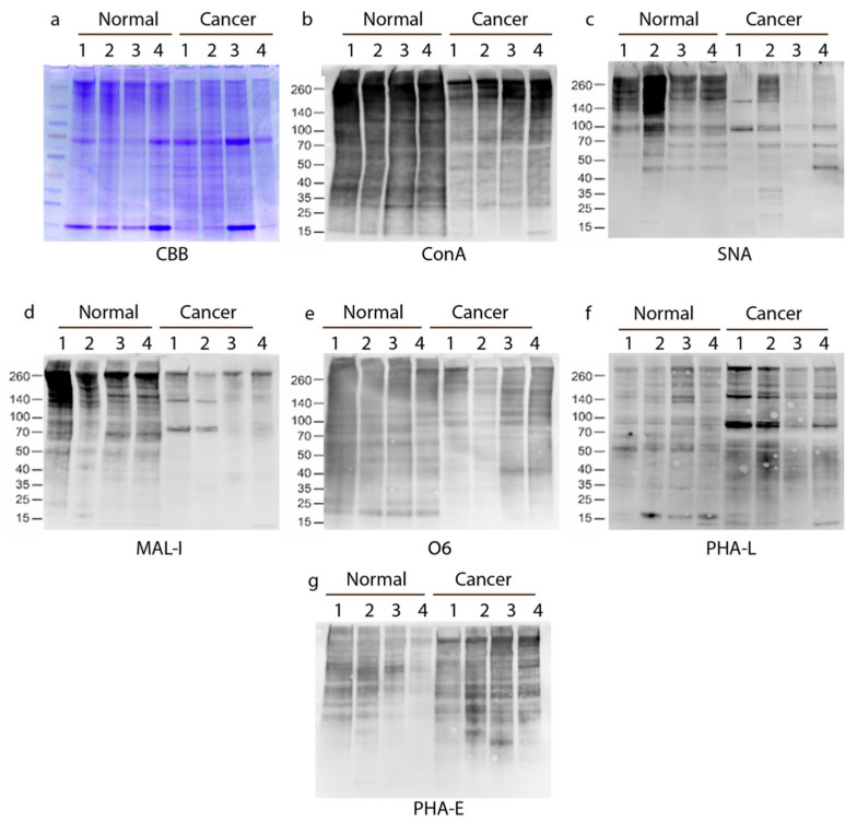
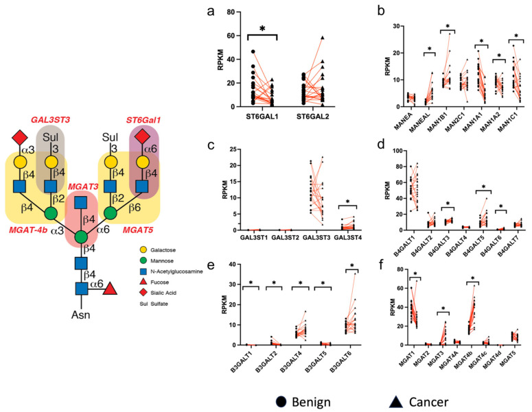
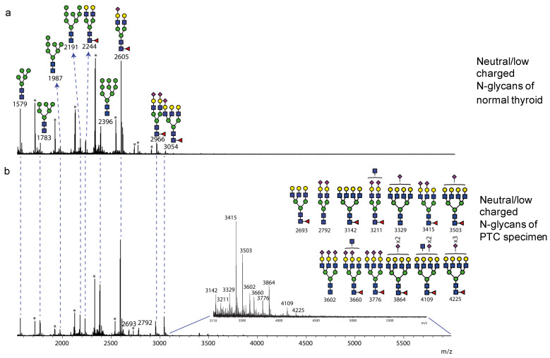
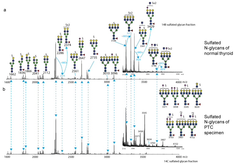
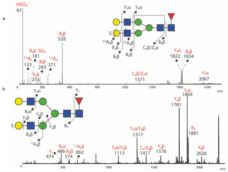
Aberrant protein glycosylation is a hallmark alteration of cancer and is highly associated with cancer progression. Papillary thyroid cancer (PTC) is the most common type of thyroid cancer, but the N-glycosylation of its glycoproteins has not been well characterized. In this work, we analyzed multiple freshly prepared PTC specimens along with paired normal tissue obtained from thyroidectomies. Glycomic analyses focused on Asn-linked (N)-glycans and employed mass spectrometry (MS), along with Western blot approaches of total solubilized materials that were examined for binding by specific lectins and a monoclonal antibody (mAb) O6, specific for 3-O-sulfated galactose residues. We observed major differences in PTC versus paired normal specimens, as PTC specimens exhibited higher levels of N-glycan branching and bisection with N-acetylglucosamine residues, consistent with RNAseq data. We also found that 3-O-sulfated galactose was present in N-glycans of multiple glycoproteins from both PTC and control specimens, as recognized by the O6 mAb and as confirmed by MS analyses. These results provide new insights into the N-glycans present in glycoproteins of thyroid cancer and context for further studies of these altered glycans as biomarkers and targets for therapeutics.

Keywords: GlcNAc bisection; RNAseq; glycosylation; lectin histochemistry; mass spectrometry; papillary thyroid cancer; sialylation; sulfation; sulfoglycomics; thyroid carcinoma; western blotting.


Conflict of interest statement

The authors declare that there are no conflicts of interest among any of the authors.

Figures

Figure 1
Figure 1
Lectin blot of normal human thyroid tissue and papillary thyroid cancer (PTC). Normal and PTC extracts of a single patient sample examined after reducing SDS-PAGE and transfer for interactions by Western blot techniques with a variety of plant lectins and the O6 mAb, whose glycan specificities are indicated in the graphic at left. Prior to gel electrophoresis, the samples were treated with or without PNGaseF (see graphic), neuraminidase A (NeuA) or neuraminidase S (NeuS) (remove sialic acid), or O-glycanase (removes core 1 O-glycans), and proteins resolved on SDS-PAGE. Gels stained with either Coomassie Brilliant Blue solution (CBB, a) or analyzed by Western/lectin blot probed with ConA (b), SNA (c), MAL-I (d), O6 VLR antibody (e), PHA-L (f), or PHA-E (g). (a) serves as a loading control for (bg). This set of gels is a representative set from 1 patient sample of 5 patients (one sample did not have enough material remaining after processing to perform these assays). Original western blots can be found at Supplementary Materials.
Figure 2
Figure 2
Lectin blot cross-comparison of four paired normal and cancerous human thyroid tissue homogenates. Tissue extracts from four of six specimens for normal and PTC were resolved on SDS-PAGE and stained with CBB (a), as in Figure 1, and in analyzed by Western/lectin blot using ConA (b), SNA (c), MAL-I (d), O6 VLR antibody (e), PHA-L (f), or PHA-E (g). (a) serves as a loading control for (bg). Original western blots can be found at Supplementary Materials.
Figure 3
Figure 3
Differential expression analysis of glycogenes. RNA sequencing data from The Cancer Genome Atlas were compared between paired normal (benign) and PTC (cancer) individual samples in units of fragments per kilobase of exon per million mapped fragments (RPKM). Graphic at left depicts the products of some of the glyco-enzyme activities. Panels are grouped by different types of glyco-enzymes, including (a) sialyltransferases, (b) mannosidases, (c) sulfotransferases, (d) β1-4galactosyltransferases, (e) β1-3galactosyltransfeases, and (f) N-acetylglucosaminyltransferases. * = p < 0.05 when comparing levels in benign and cancer tissues. For all panels, n = 20 for each specified benign and cancer group.
Figure 4
Figure 4
Neutral N-glycan profile of normal thyroid tissue and PTC from matching patient. Neutral N-glycans released from thyroid homogenates by PNGase F following mild permethylation and fractionation by MAX columns. The data were acquired by MALDI-TOF MS in the positive ion mode. The medial mass-to-charge range is shown from m/z 1550 to 6000. The ions corresponding to fully permethylated glycan structures are labelled with its molecular mass, putative glycan structures are also assigned to each labelled mass. Signals with identical molecular mass are matched by dash lines between the spectra. Signals not corresponding to fully permethylated N-glycan structures are noted with an asterisk *. (a) Neutral N-glycan profile of thyroid normal tissue; (b) Neutral N-glycan profile of PTC.
Figure 5
Figure 5
Sulfo N-glycan profile of normal thyroid tissue and PTC from matching patient. Sulfated N-glycans from the charged glycan fraction were PNGase F released, permethylated, and fractionated by MAX column. The MS profile spectra were acquired in the negative ion mode MALDI-TOF MS. The medial range is m/z 1600 to m/z 4000 as shown. The inset medial range shown is from m/z 3110 to 4200. The fully permethylated mono-sulfated N-glycans are labelled with black number and their putative structures are also assigned with a “S” symbol above the cartoon to represent the presence of a sulfo group. Ions corresponding to putatively di-sulfated N-glycans are labelled with blue numbers and the +88Da mass adduct is also labelled. Signals with identical molecular mass for glycan structures are matched between spectra with dash lines. (a) Sulfo N-glycan profile of normal thyroid tissue; (b) Sulfo N-glycan profile of PTC.
Figure 6
Figure 6
MS/MS fragmentation analyses for m/z 2286 and 2332. In-source MS/MS fragmentation analyses were performed by MALDI TOF/TOF for selected ions. Signals for the corresponding fragment ions are labelled with their m/z value. The parent ion cartoons are shown with the arrowed lines and numbers representing the corresponding fragments. The symbol “S” is labelled on the glycan cartoon to represent sulfation. The fragment ion assignments are based on the Domon-Costello fragmentation pathways for glycans [56]. (a) MS/MS fragment spectrum of m/z 2286 in the negative mode; (b) MS/MS fragment spectrum of m/z 2332 in the positive mode.

Similar articles

Cited by

References

    1. Pinho S.S., Reis C.A. Glycosylation in cancer: Mechanisms and clinical implications. Nat. Rev. Cancer. 2015;15:540–555. doi: 10.1038/nrc3982. - DOI - PubMed
    1. Stowell S.R., Ju T., Cummings R.D. Protein glycosylation in cancer. Annu. Rev. Pathol. 2015;10:473–510. doi: 10.1146/annurev-pathol-012414-040438. - DOI - PMC - PubMed
    1. Oliveira-Ferrer L., Legler K., Milde-Langosch K. Role of protein glycosylation in cancer metastasis. Semin. Cancer Biol. 2017;44:141–152. doi: 10.1016/j.semcancer.2017.03.002. - DOI - PubMed
    1. Bellis S.L., Reis C.A., Varki A., Kannagi R., Stanley P. Glycosylation Changes in Cancer. In: Varki A., Cummings R.D., Esko J.D., Stanley P., Hart G.W., Aebi M., Mohnen D., Kinoshita T., Packer N.H., Prestegard J.H., et al., editors. Essentials of Glycobiology. 4th ed. Cold Spring Harbor Laboratory Press; Cold Spring Harbor, NY, USA: 2022. pp. 631–644.
    1. Munkley J., Elliott D.J. Hallmarks of glycosylation in cancer. Oncotarget. 2016;7:35478–35489. doi: 10.18632/oncotarget.8155. - DOI - PMC - PubMed

LinkOut - more resources