. 2024 Nov 23;14(12):1492.
doi: 10.3390/biom14121492.

Diabetic Glycation of Human Serum Albumin Affects Its Immunogenicity

Affiliations

Diabetic Glycation of Human Serum Albumin Affects Its Immunogenicity

Cresci-Anne C C Croes et al. Biomolecules. .

Abstract

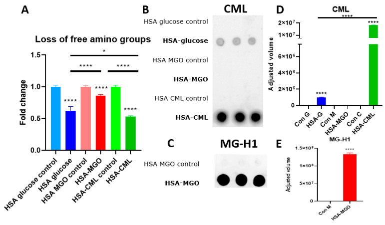
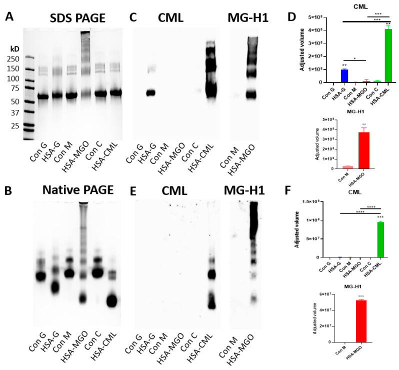
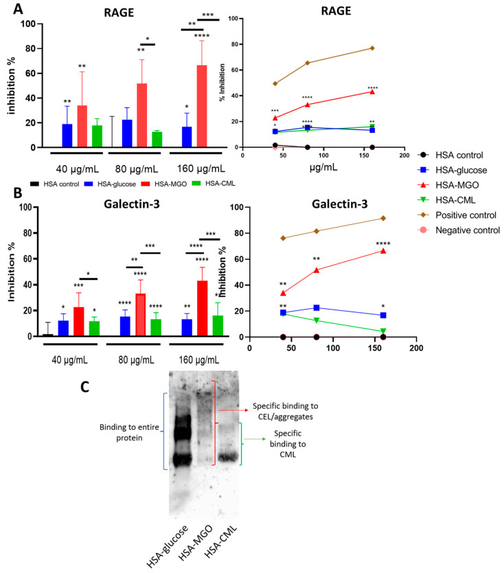
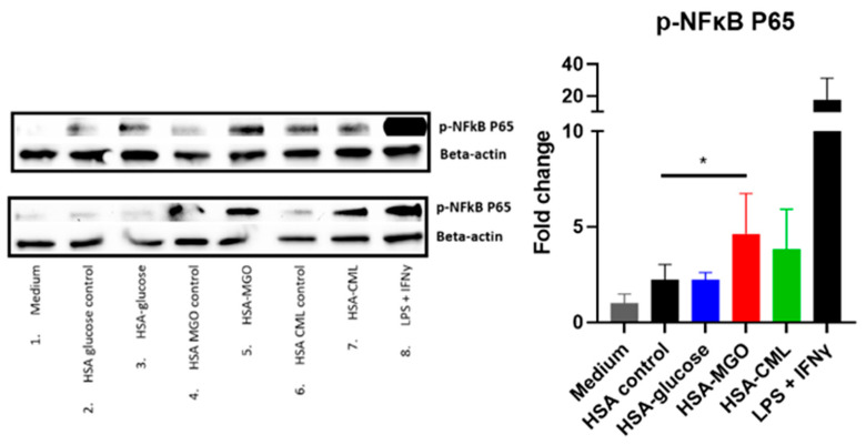
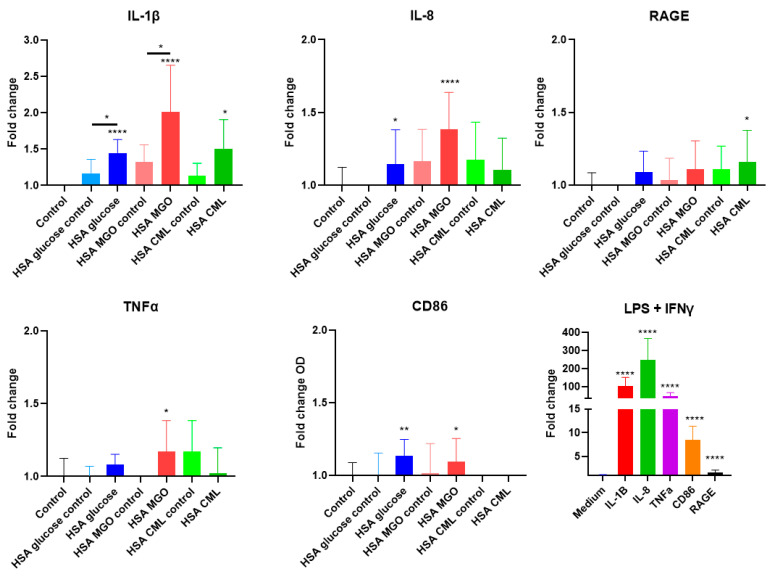
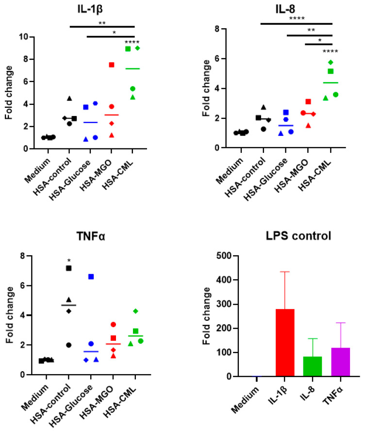
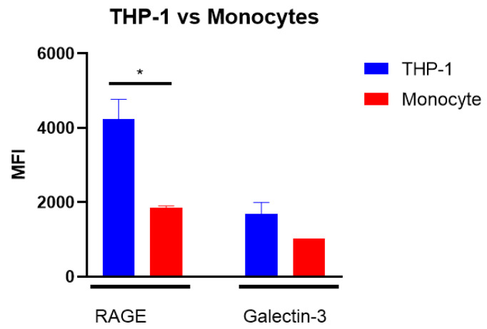
Advanced glycation end-products (AGEs) are products of a non-enzymatic reaction between amino acids and reducing sugars. Glycated human serum albumin (HSA) increases in diabetics as a consequence of elevated blood glucose levels and glycating metabolites like methylglyoxal (MGO). The impact of different types of glycation on the immunomodulatory properties of HSA is poorly understood and is studied here. HSA was glycated with D-glucose, MGO, or glyoxylic acid (CML). Glycation-related biochemical changes were characterized using various biochemical methods. The binding of differentially glycated HSA to AGE receptors was determined with inhibition ELISAs, and the impact on inflammatory markers in macrophage cell line THP-1 and adherent monocytes isolated from human peripheral blood mononuclear cells (PBMCs) was studied. All glycation methods led to unique AGE profiles and had a distinct impact on protein structure. Glycation resulted in increased binding of HSA to the AGE receptors, with MGO modification showing the highest binding, followed by glucose and, lastly, CML. Additionally, modification of HSA with MGO led to the increased expression of pro-inflammatory markers in THP-1 macrophages and enhanced phosphorylation of NF-κB p65. The same pattern, although less prominent, was observed for HSA glycated with glucose and CML, respectively. An increase in pro-inflammatory markers was also observed in PBMC-derived monocytes exposed to all glycated forms of HSA, although HSA-CML led to a significantly higher inflammatory response. In conclusion, the type of HSA glycation impacts immune functional readouts with potential relevance for diabetes.

Keywords: AGEs; HSA; RAGE; diabetes; inflammation; macrophages; methylglyoxal; receptors.


Conflict of interest statement

The authors declare no conflicts of interest.

Figures

Figure 1
Figure 1
Confirmation of glycation of human serum albumin. (A) Loss of amino groups measured with OPA assay. (B,C) The presence of CML or MG-H1 was measured with dot blot and (D,E) quantified data of dot blot. Data presented as triplicate, 30 μg per dot. Data shown as mean ± SD of triplicate wells. Significant differences were analyzed with the Student’s t-test (GraphPad Prism): * p < 0.05, **** p < 0.0001.
Figure 2
Figure 2
Effect of glycation on the HSA protein. A total of 8 μg of glycated HSA and non-glycated HSA controls were analyzed via SDS-PAGE (A) and NATIVE-PAGE (B). Gels were visualized with Coomassie brilliant blue staining. Additionally, a Western blot was performed after denaturing, and Native Gel electrophoresis and tested with anti-CML and anti-MG-H1 antibodies, and data were quantified for both SDS (C,D) and NATIVE (E,F) Gels. Data were analyzed using the ImageLab software by Biorad. Significant differences analyzed with the Student’s t-test (GraphPad Prism); * p < 0.05, ** p < 0.01, *** p < 0.001, **** p < 0.0001.
Figure 3
Figure 3
Binding of glycated HSA to AGE receptors. Glycated HSA and non-glycated HSA analyzed via inhibition ELISA for binding efficiency against (A) RAGE and (B) Galectin-3. Glycated soy and ovalbumin were used as positive and negative controls. Western blot against RAGE receptor showing to which glycated HSA RAGE binds (C). Data shown as mean ± SD of duplicate wells and are representative of three individual experiments. Significant differences analyzed with the Student’s t-test (GraphPad Prism); * p < 0.05, ** p < 0.01, *** p < 0.001, **** p < 0.0001.
Figure 4
Figure 4
Phosphorylated NF-κB P65 protein expression in response to treatment with glycated HSA in THP-1 macrophages. THP-1-derived macrophages were stimulated with either 100 μg/mL of glycated HSA or non-glycated HSA for 10 min, followed by Western blot analyses for phosphorylated NF-κB P65 protein expression. Stimulation with LPS and IFNy was used as positive control. Data shown as mean ± SD of duplicate experiments. Western blot data quantified and analyzed using ImageLab software from Biorad. HSA controls for all 3 glycations were pooled together for quantification. Significant differences analyzed with the Student’s t-test (GraphPad Prism); * p < 0.05.
Figure 5
Figure 5
The effect of stimulation with glycated HSA on pro-inflammatory markers in THP-1 macrophages. THP-1 macrophages were stimulated with 100 μg/mL of either glycated HSA or non-glycated HSA for either 8 h (HSA–MGO) or for 24 h (HSA–glucose and HSA–CML), followed by qPCR analyses. Data shown as mean ± SD of triplicate wells and are representative of at least two individual experiments. LPS and IFNy were used as a positive control, as shown in a separate graph. Buffer controls are shown in Supplementary Figure S6. Significant differences analyzed with the Student’s t-test (GraphPad Prism); * p < 0.05, ** p < 0.01, **** p < 0.0001.
Figure 6
Figure 6
Effect of stimulation with glycated HSA on primary monocytes. PBMC-derived monocytes from 4 donors were stimulated with 100 μg/mL of either glycated HSA or non-glycated HSA for 3 h, followed by qPCR analysis of IL-1β, IL-8, and TNFα, and positive control LPS + IFNy showing that donors monocyte respond well to inflammatory stimuli. Data shown as mean ± SD of duplicate wells. Combined data of all donors, Significant differences analyzed with One-way ANOVA (GraphPad Prism); * p < 0.05, ** p < 0.01, **** p < 0.0001.
Figure 7
Figure 7
Expression of receptors on THP-1 macrophages and primary adherent monocytes. THP-1 macrophages and PBMC-derived adherent monocytes were analyzed via a Flow cytometer. Significant differences analyzed with the Student’s t-test (GraphPad Prism); * p < 0.05.

Similar articles

References

    1. Khan M.A.B., Hashim M.J., King J.K., Govender R.D., Mustafa H., Kaabi J.A. Epidemiology of Type 2 Diabetes—Global Burden of Disease and Forecasted Trends. J. Epidemiol. Glob. Health. 2019;10:107–111. doi: 10.2991/jegh.k.191028.001. - DOI - PMC - PubMed
    1. Qiu H.-Y., Hou N.-N., Shi J.-F., Liu Y.-P., Kan C.-X., Han F., Sun X.-D. Comprehensive overview of human serum albumin glycation in diabetes mellitus. World J. Diabetes. 2021;12:1057–1069. doi: 10.4239/wjd.v12.i7.1057. - DOI - PMC - PubMed
    1. Indurthi V.S.K., Leclerc E., Vetter S.W. Interaction between glycated serum albumin and AGE-receptors depends on structural changes and the glycation reagent. Arch. Biochem. Biophys. 2012;528:185–196. doi: 10.1016/j.abb.2012.10.002. - DOI - PubMed
    1. Kumari N., Bandyopadhyay D., Kumar V., Venkatesh D.B., Prasad S., Prakash S., Krishnaswamy P.R., Balaram P., Bhat N. Glycation of albumin and its implication in Diabetes: A comprehensive analysis using mass spectrometry. Clin. Chim. Acta. 2021;520:108–117. doi: 10.1016/j.cca.2021.06.001. - DOI - PubMed
    1. Rabbani G., Ahn S.N. Structure, enzymatic activities, glycation and therapeutic potential of human serum albumin: A natural cargo. Int. J. Biol. Macromol. 2019;123:979–990. doi: 10.1016/j.ijbiomac.2018.11.053. - DOI - PubMed

MeSH terms

LinkOut - more resources