. 2024 Nov 26;14(12):1506.
doi: 10.3390/biom14121506.

Steroids and Malignancy Increase Local Heparanase and Decrease Markers of Osteoblast Activity in Bone Tissue Microcirculation

Affiliations

Steroids and Malignancy Increase Local Heparanase and Decrease Markers of Osteoblast Activity in Bone Tissue Microcirculation

Keren Asayag et al. Biomolecules. .

Abstract

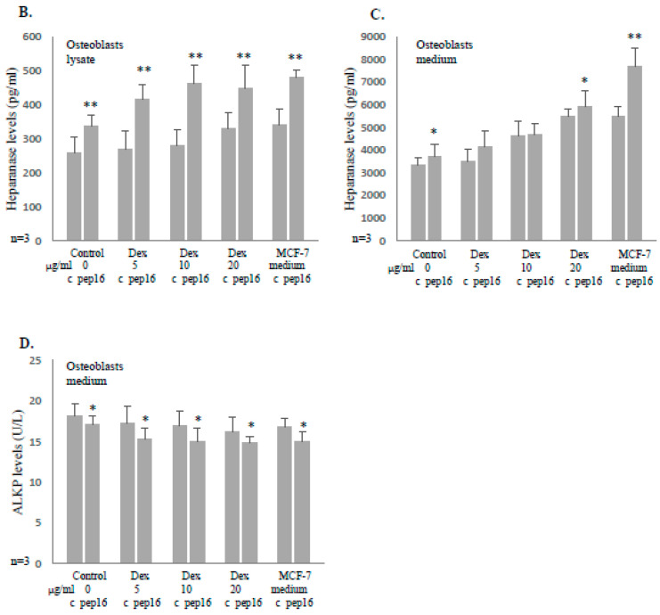
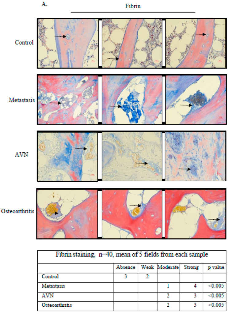
Bone metastasis and steroids are known to activate the coagulation system and induce osteoporosis, pathological bone fractures, and bone pain. Heparanase is a protein known to enhance the hemostatic system and to promote angiogenesis, metastasis, and inflammation. The objective of the present study was to evaluate the effects of steroids and malignancy on the coagulation factors and osteoblast activity in the bone tissue. The effects of dexacort and malignant medium were evaluated in osteoblasts derived from human bone marrow mesenchymal stem cells and human umbilical vein endothelial cells (HUVECs). The bones of mice treated with dexacort for 1 month were studied. Bone biopsies of ten patients with bone metastasis, ten with steroid-induced avascular necrosis (AVN), and ten with osteoarthritis were compared to ten controls. We found that dexacort and malignant medium significantly increased the heparanase levels in osteoblasts and HUVECs and decreased the levels of alkaline phosphatase (ALKP). Peptide 16AC, derived from heparanase, which interacts with tissue factor (TF), further increased the effect, while peptide 6, which inhibits interactions between heparanase and TF, reversed the effect in these cells. The bone microcirculation of mice treated with dexacort exhibited significantly higher levels of heparanase, TF, TF pathway inhibitor (TFPI), TFPI-2, thrombin, and syndecan-1, but reduced levels of osteocalcin and ALKP. The pathological human bone biopsies' microcirculation exhibited significantly dilated blood vessels and higher levels of heparanase, TF, TFPI, TFPI-2, and fibrin. In summary, steroids and malignancy increased the activation of the coagulation system in the bone microcirculation and reduced the osteoblast activity. Heparanase inhibitors should be further investigated to attenuate bone fractures and pain.

Keywords: bone; heparanase; malignancy; microcirculation; steroids; thrombosis.


Conflict of interest statement

The authors declare no conflicts of interest.

Figures

Figure 1
Figure 1
Dexacort or medium of malignant cells increases heparanase expression and reduces ALKP in osteoblast cells. Human MSCs were isolated from human BM pellet and differentiated to osteoblasts with osteogenic medium, as described in the Methods section. After 3 weeks, the osteoblasts were incubated for 48 h under the following conditions: 1. control (1000 μL osteogenic medium); 2. dexacort in increasing concentrations equivalent to therapeutic doses (2.5 µg/mL, 5 µg/mL, 10 µg/mL) and osteogenic medium; and 3. osteogenic medium (1000 µL) derived from overnight incubation with three malignant cell lines (MCF-7—human breast carcinoma, A549—human lung adenocarcinoma, and MDA-231—human breast carcinoma). (A) Immunohistochemistry staining revealed that osteoblasts incubated with dexacort or medium from malignant cells showed a significantly higher level of heparanase. The contiguous table shows the indicated protein staining intensity in the cells. Significance was determined by the Mann–Whitney U test. Representative images were visualized through ×10 magnitude, with 0.82 MDC objective lens, captured with a Nikon E995 digital camera (Nikon, Tokyo, Japan), and processed with Adobe Photoshop software (Adobe Systems, San Jose, CA, USA). The heparanase levels in the mediums and lysates were tested by ELISA assay, as described in the Methods. The results show increased heparanase levels in both the osteoblast lysates and medium after incubation with dexacort (B,C) or medium from malignant cells (D,E). ALKP levels by spectrophotometry analysis that indicate the osteoblast activity showed that dexacort and malignant medium reduced the levels of ALKP significantly (F). The effect of malignant medium was compared to the level of ALKP in the medium before incubation with osteoblasts (light gray). Significance was determined by the Mann–Whitney U test. All the assays were performed in triplicate. Results represent median and range of three experiments. * p < 0.05, ** p < 0.005.
Figure 1
Figure 1
Dexacort or medium of malignant cells increases heparanase expression and reduces ALKP in osteoblast cells. Human MSCs were isolated from human BM pellet and differentiated to osteoblasts with osteogenic medium, as described in the Methods section. After 3 weeks, the osteoblasts were incubated for 48 h under the following conditions: 1. control (1000 μL osteogenic medium); 2. dexacort in increasing concentrations equivalent to therapeutic doses (2.5 µg/mL, 5 µg/mL, 10 µg/mL) and osteogenic medium; and 3. osteogenic medium (1000 µL) derived from overnight incubation with three malignant cell lines (MCF-7—human breast carcinoma, A549—human lung adenocarcinoma, and MDA-231—human breast carcinoma). (A) Immunohistochemistry staining revealed that osteoblasts incubated with dexacort or medium from malignant cells showed a significantly higher level of heparanase. The contiguous table shows the indicated protein staining intensity in the cells. Significance was determined by the Mann–Whitney U test. Representative images were visualized through ×10 magnitude, with 0.82 MDC objective lens, captured with a Nikon E995 digital camera (Nikon, Tokyo, Japan), and processed with Adobe Photoshop software (Adobe Systems, San Jose, CA, USA). The heparanase levels in the mediums and lysates were tested by ELISA assay, as described in the Methods. The results show increased heparanase levels in both the osteoblast lysates and medium after incubation with dexacort (B,C) or medium from malignant cells (D,E). ALKP levels by spectrophotometry analysis that indicate the osteoblast activity showed that dexacort and malignant medium reduced the levels of ALKP significantly (F). The effect of malignant medium was compared to the level of ALKP in the medium before incubation with osteoblasts (light gray). Significance was determined by the Mann–Whitney U test. All the assays were performed in triplicate. Results represent median and range of three experiments. * p < 0.05, ** p < 0.005.
Figure 2
Figure 2
Heparanase expression was increased and ALKP decreased in osteoblast cells when incubated with the heparanase procoagulant peptide 16AC. MSCs were isolated from human BM pellet and differentiated to osteoblasts with osteogenic medium. After 3 weeks, the osteoblasts were incubated for 48 h under the following conditions: 1. control (1000 μL osteogenic medium); 2. dexacort in increasing concentrations equivalent to therapeutic doses (5 µg/mL, 10 µg/mL, and 20 µg/mL) in osteogenic medium; and 3. osteogenic medium (1000 µL) derived from overnight incubation with malignant cell line (MCF-7). Peptide 16AC (designated 16, 5 μg/mL) was added to half of the plates. (A) Immunohistochemistry staining. The results revealed a significant increase in heparanase expression in osteoblast cells incubated with peptide 16AC and dexacort or medium from malignant cells. The contiguous table shows the indicated protein staining intensity in the cells. Significance was determined by the Mann–Whitney U test. Representative images were visualized through ×50 magnitude, with 0.82 MDC objective lens, captured with a Nikon E995 digital camera (Nikon, Tokyo, Japan), and processed with Adobe Photoshop software (Adobe Systems, San Jose, CA, USA). (B,C) The heparanase levels in mediums and lysates were tested by ELISA assay. The results show that peptide 16AC has an additive effect on the heparanase levels in osteoblast cell lysate and medium incubated with dexacort or malignant cells medium. All the assays were performed in triplicate. The results represent the median and range of three experiments. (D) The ALKP level was measured in the medium of the above-described experiment. The results indicate that peptide 16AC has an additive effect on decreasing the ALKP levels in osteoblast cells incubated with dexacort or malignant cells medium. The results represent the median and range of three experiments. * p < 0.05, ** p < 0.005.
Figure 2
Figure 2
Heparanase expression was increased and ALKP decreased in osteoblast cells when incubated with the heparanase procoagulant peptide 16AC. MSCs were isolated from human BM pellet and differentiated to osteoblasts with osteogenic medium. After 3 weeks, the osteoblasts were incubated for 48 h under the following conditions: 1. control (1000 μL osteogenic medium); 2. dexacort in increasing concentrations equivalent to therapeutic doses (5 µg/mL, 10 µg/mL, and 20 µg/mL) in osteogenic medium; and 3. osteogenic medium (1000 µL) derived from overnight incubation with malignant cell line (MCF-7). Peptide 16AC (designated 16, 5 μg/mL) was added to half of the plates. (A) Immunohistochemistry staining. The results revealed a significant increase in heparanase expression in osteoblast cells incubated with peptide 16AC and dexacort or medium from malignant cells. The contiguous table shows the indicated protein staining intensity in the cells. Significance was determined by the Mann–Whitney U test. Representative images were visualized through ×50 magnitude, with 0.82 MDC objective lens, captured with a Nikon E995 digital camera (Nikon, Tokyo, Japan), and processed with Adobe Photoshop software (Adobe Systems, San Jose, CA, USA). (B,C) The heparanase levels in mediums and lysates were tested by ELISA assay. The results show that peptide 16AC has an additive effect on the heparanase levels in osteoblast cell lysate and medium incubated with dexacort or malignant cells medium. All the assays were performed in triplicate. The results represent the median and range of three experiments. (D) The ALKP level was measured in the medium of the above-described experiment. The results indicate that peptide 16AC has an additive effect on decreasing the ALKP levels in osteoblast cells incubated with dexacort or malignant cells medium. The results represent the median and range of three experiments. * p < 0.05, ** p < 0.005.
Figure 3
Figure 3
Heparanase inhibitory peptide 6 attenuates the increase in heparanase and reduction in ALKP induced by dexacort and medium from malignant cells in osteoblast cells. MSCs were isolated from human BM pellet and differentiated to osteoblasts with osteogenic medium. After 3 weeks, osteoblasts were incubated for 48 h under the following conditions: 1. control (1000 μL osteogenic medium); 2. dexacort in increasing concentrations equivalent to therapeutic doses (5 µg/mL, 10 µg/mL, and 20 µg/mL) and osteogenic medium; and 3. osteogenic medium (1000 µL) was derived from overnight incubation with malignant cells (A,B). Peptide 6 (50 μg/mL) was added to half of the plates. (A,B) Immunohistochemistry staining and ELISA. The results revealed that peptide 6 attenuated the heparanase increase induced by dexacort and malignant medium in osteoblast cells. Contiguous table shows the indicated protein staining intensity in the cells. Significance was determined by the Mann–Whitney U test. Representative images were visualized through ×50 magnitude, with 0.82 MDC objective lens, captured with a Nikon E995 digital camera (Nikon, Tokyo, Japan), and processed with Adobe Photoshop software (Adobe Systems, San Jose, CA, USA). (C) ALKP level. The results indicate that peptide 6 reduced the ALKP level when added to osteoblast cells incubated with dexacort or medium from malignant cells. The results represent the median and range of three experiments.* p < 0.05, ** p < 0.005.
Figure 3
Figure 3
Heparanase inhibitory peptide 6 attenuates the increase in heparanase and reduction in ALKP induced by dexacort and medium from malignant cells in osteoblast cells. MSCs were isolated from human BM pellet and differentiated to osteoblasts with osteogenic medium. After 3 weeks, osteoblasts were incubated for 48 h under the following conditions: 1. control (1000 μL osteogenic medium); 2. dexacort in increasing concentrations equivalent to therapeutic doses (5 µg/mL, 10 µg/mL, and 20 µg/mL) and osteogenic medium; and 3. osteogenic medium (1000 µL) was derived from overnight incubation with malignant cells (A,B). Peptide 6 (50 μg/mL) was added to half of the plates. (A,B) Immunohistochemistry staining and ELISA. The results revealed that peptide 6 attenuated the heparanase increase induced by dexacort and malignant medium in osteoblast cells. Contiguous table shows the indicated protein staining intensity in the cells. Significance was determined by the Mann–Whitney U test. Representative images were visualized through ×50 magnitude, with 0.82 MDC objective lens, captured with a Nikon E995 digital camera (Nikon, Tokyo, Japan), and processed with Adobe Photoshop software (Adobe Systems, San Jose, CA, USA). (C) ALKP level. The results indicate that peptide 6 reduced the ALKP level when added to osteoblast cells incubated with dexacort or medium from malignant cells. The results represent the median and range of three experiments.* p < 0.05, ** p < 0.005.
Figure 4
Figure 4
Peptide 6 reduces heparanase level and increases ALKP level induced by dexacort in HUVECs. (A) HUVECs at the 5th passage were incubated for 48 h under the following conditions: 1. control (1000 μL M-199 medium); and 2. dexacort in increasing concentrations equivalent to therapeutic doses (2.5 µg/mL, 5 µg/mL, and 10 µg/mL) and M-199 medium. Heparanase inhibitory peptide 6 (50 μg/mL) was added to half of the plates. The results show that dexacort increased and peptide 6 reduced the secretion of heparanase into the medium when added to HUVECs. (B) The ALKP level was evaluated in the above-described experiment. While dexacort increased, peptide 6 reduced the level of ALKP in the medium when added to HUVECs. All the assays were performed in triplicates. The results represent the median and range of three experiments.* p < 0.05, ** p < 0.005.
Figure 5
Figure 5
Dexacort increases the levels of coagulation parameters and decreases the markers of osteoblast activity in mice bone tissue microcirculation. (A) Immunohistochemical staining was performed in proximal bone tissue of mice. The control group (n = 6) received water without any drug for one month. The treatment group (n = 6) received water with the addition of dexacort (equivalent to a human therapeutic dose of 12 mg/day) for 1 month. The water was replaced every 48 h. At the end of the trial, the mice were sacrificed and the posterior proximal leg bones harvested. Slides were stained to the following five coagulation parameters: heparanase, TF, TFPI, TFPI-2, and thrombin, to two heparan sulfate proteoglycans that heparanase degrades; syndecan-1 and perlecan, in addition to osteocalcin, which is a marker of osteoblast activity. A significant increase in all the coagulation parameters, including syndecan-1, and a significant decrease in osteocalcin were observed in bone tissue of the treatment mice group as compared to the control mice group, mainly in the bone microcirculation and bone marrow sinusoids. The contiguous table shows the indicated protein staining intensity in the cells. Significance was determined by the Mann–Whitney U test. Representative images were visualized through ×50 magnitude, with a 0.82 MDC objective lens, captured with a Nikon E995 digital camera (Nikon, Tokyo, Japan), and processed with Adobe Photoshop software (Adobe Systems, San Jose, CA, USA) (AG). No effect was found on the level of perlecan (H). In addition, bone samples were pulverized with a hammer in liquid nitrogen, extractions were centrifuged at 13,000× g for 15 min, and the supernatant was evaluated. The heparanase levels determined by ELISA were significantly higher and the ALKP levels were significantly lower in the study group as compared to the controls (I,J). All the assays were performed in triplicate. The results represent the median and range. The Mann–Whitney U test was used. ** p < 0.005.
Figure 5
Figure 5
Dexacort increases the levels of coagulation parameters and decreases the markers of osteoblast activity in mice bone tissue microcirculation. (A) Immunohistochemical staining was performed in proximal bone tissue of mice. The control group (n = 6) received water without any drug for one month. The treatment group (n = 6) received water with the addition of dexacort (equivalent to a human therapeutic dose of 12 mg/day) for 1 month. The water was replaced every 48 h. At the end of the trial, the mice were sacrificed and the posterior proximal leg bones harvested. Slides were stained to the following five coagulation parameters: heparanase, TF, TFPI, TFPI-2, and thrombin, to two heparan sulfate proteoglycans that heparanase degrades; syndecan-1 and perlecan, in addition to osteocalcin, which is a marker of osteoblast activity. A significant increase in all the coagulation parameters, including syndecan-1, and a significant decrease in osteocalcin were observed in bone tissue of the treatment mice group as compared to the control mice group, mainly in the bone microcirculation and bone marrow sinusoids. The contiguous table shows the indicated protein staining intensity in the cells. Significance was determined by the Mann–Whitney U test. Representative images were visualized through ×50 magnitude, with a 0.82 MDC objective lens, captured with a Nikon E995 digital camera (Nikon, Tokyo, Japan), and processed with Adobe Photoshop software (Adobe Systems, San Jose, CA, USA) (AG). No effect was found on the level of perlecan (H). In addition, bone samples were pulverized with a hammer in liquid nitrogen, extractions were centrifuged at 13,000× g for 15 min, and the supernatant was evaluated. The heparanase levels determined by ELISA were significantly higher and the ALKP levels were significantly lower in the study group as compared to the controls (I,J). All the assays were performed in triplicate. The results represent the median and range. The Mann–Whitney U test was used. ** p < 0.005.
Figure 6
Figure 6
Malignancy, steroids and inflammation induce coagulation activation in human bone microcirculation. After obtaining written informed consent, the bone biopsies of 40 patients were studied: ten cases of bone metastasis of carcinoma origin, ten cases of avascular necrosis (AVN) of the femur head due to steroids, and ten cases of osteoarthritis of the femur head. These biopsies were taken at the beginning of femur surgery. Ten cases of diffuse large cell lymphoma without bone or bone marrow involvement, which formed the control group, were harvested during the bone marrow biopsy procedure during staging. There was no difference in gender, age, concomitant illnesses, or anti-aggregant use in the four groups. Data are presented in Table 1. The bone biopsies were studied using specific staining to fibrin with Martin Scarlet Blue (MSB) and immune-staining to heparanase, TF, TFPI, and TFPI-2. We found that the blood vessels in the study group were intensely dilated in the bone tissue and bone marrow sinuses ((A,B), indicated by black arrows). In MSB, staining fibrin is blue and erythrocytes are yellow. Fibrin was present at a significantly higher level in the study group microcirculation as compared to the control. As the staining of MSB is specific to fibrin and does not stain fibrinogen, the fibrin found in the microcirculation represents micro-thrombi (A). Increased levels of heparanase, TF, TFPI, and TFPI-2 in the study group, as compared to the control, were found. Increased levels of heparanase and TF may induce activation of the coagulation system (B). Contiguous tables show the indicated protein staining intensity in cells. Significance was determined by Mann-Whitney U test. Representative images were visualized through x10 magnitude, with a 0.82 MDC objective lens, captured with a Nikon E995 digital camera (Nikon, Tokyo, Japan), and processed with Adobe Photoshop software (Adobe Systems, San Jose, CA, USA).
Figure 6
Figure 6
Malignancy, steroids and inflammation induce coagulation activation in human bone microcirculation. After obtaining written informed consent, the bone biopsies of 40 patients were studied: ten cases of bone metastasis of carcinoma origin, ten cases of avascular necrosis (AVN) of the femur head due to steroids, and ten cases of osteoarthritis of the femur head. These biopsies were taken at the beginning of femur surgery. Ten cases of diffuse large cell lymphoma without bone or bone marrow involvement, which formed the control group, were harvested during the bone marrow biopsy procedure during staging. There was no difference in gender, age, concomitant illnesses, or anti-aggregant use in the four groups. Data are presented in Table 1. The bone biopsies were studied using specific staining to fibrin with Martin Scarlet Blue (MSB) and immune-staining to heparanase, TF, TFPI, and TFPI-2. We found that the blood vessels in the study group were intensely dilated in the bone tissue and bone marrow sinuses ((A,B), indicated by black arrows). In MSB, staining fibrin is blue and erythrocytes are yellow. Fibrin was present at a significantly higher level in the study group microcirculation as compared to the control. As the staining of MSB is specific to fibrin and does not stain fibrinogen, the fibrin found in the microcirculation represents micro-thrombi (A). Increased levels of heparanase, TF, TFPI, and TFPI-2 in the study group, as compared to the control, were found. Increased levels of heparanase and TF may induce activation of the coagulation system (B). Contiguous tables show the indicated protein staining intensity in cells. Significance was determined by Mann-Whitney U test. Representative images were visualized through x10 magnitude, with a 0.82 MDC objective lens, captured with a Nikon E995 digital camera (Nikon, Tokyo, Japan), and processed with Adobe Photoshop software (Adobe Systems, San Jose, CA, USA).
Figure 7
Figure 7
Schematic summarization. (Left) The blood supply by the nutrient vessels to the bone and bone marrow is the same, thus both can be considered one organ. (Right) Steroids and malignant milieu in osteoblasts and endothelial cells induce upregulation of proteins involved in microcirculation thrombosis and downregulation of proteins involved in bone remodeling. Heparanase up-regulates itself using a positive feedback mechanism. Inhibition of heparanase restores the hemostatic balance and osteoblast activity.

Similar articles

Cited by

References

    1. McCarthy I. The physiology of bone blood flow: A review. J. Bone Jt. Surg. Am. 2006;88((Suppl. S3)):4–9. doi: 10.2106/00004623-200611001-00002. - DOI - PubMed
    1. Marenzana M., Arnett T.R. The Key Role of the Blood Supply to Bone. Bone Res. 2013;1:203–215. doi: 10.4248/BR201303001. - DOI - PMC - PubMed
    1. Del Fattore A., Capannolo M., Rucci N. Bone and bone marrow: The same organ. Arch. Biochem. Biophys. 2010;503:28–34. doi: 10.1016/j.abb.2010.07.020. - DOI - PubMed
    1. Feingold K.R., Anawalt B., Blackman M.R., Boyce A., Chrousos G., Corpas E., de Herder W.W., Dhatariya K., Dungan K., Hofland J., et al., editors. Endotext. MDText.com, Inc.; South Dartmouth, MA, USA: 2000. An Overview of Glucocorticoid-Induced Osteoporosis. - PubMed
    1. Legakis I., Syrigos K. Osteoporosis in patients with breast and prostate cancer: Effect of disease and treatment modalities. Endocr. Metab. Immune Disord. Drug Targets. 2013;13:168–174. doi: 10.2174/18715303112129990027. - DOI - PubMed

MeSH terms

LinkOut - more resources