Review
. 2024 Dec 9;14(12):1569.
doi: 10.3390/biom14121569.

Organ-on-a-Chip Models-New Possibilities in Experimental Science and Disease Modeling

Affiliations
Review

Organ-on-a-Chip Models-New Possibilities in Experimental Science and Disease Modeling

Bartłomiej Wysoczański et al. Biomolecules. .

Abstract

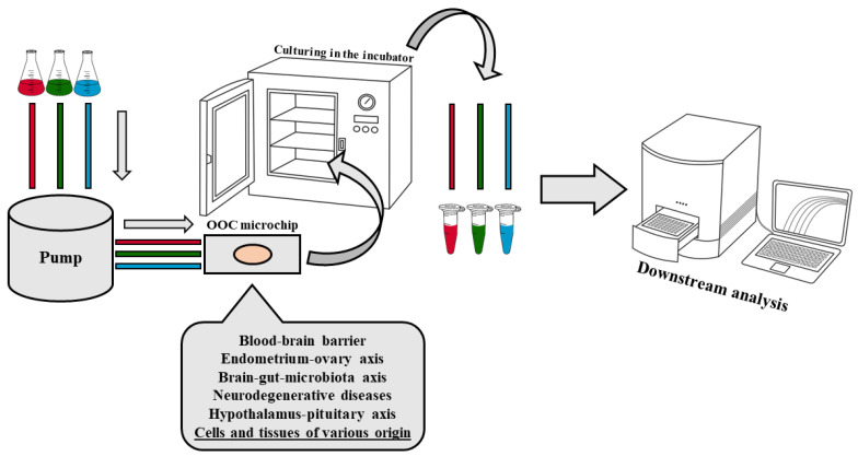
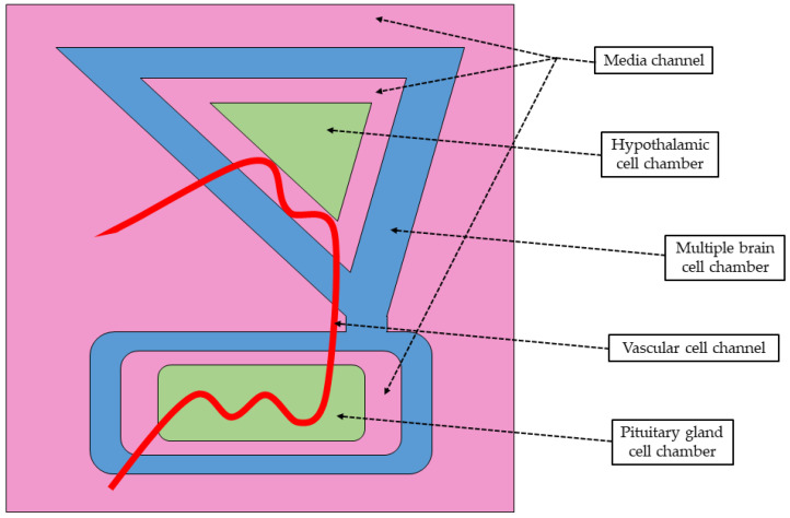
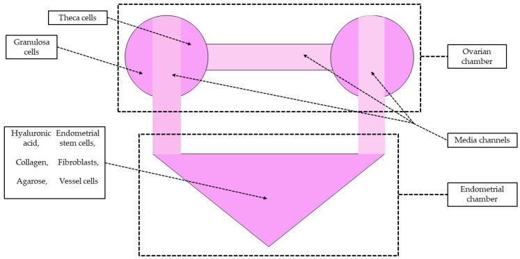
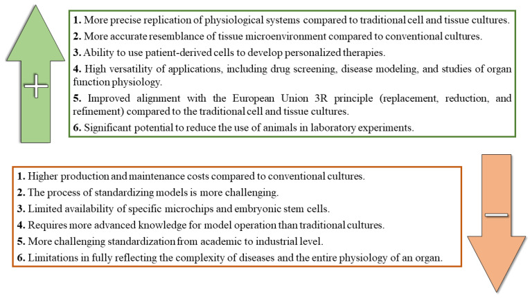
'Organ-on-a-chip' technology is a promising and rapidly evolving model in biological research. This innovative microfluidic cell culture device was created using a microchip with continuously perfused chambers, populated by living cells arranged to replicate physiological processes at the tissue and organ levels. By consolidating multicellular structures, tissue-tissue interfaces, and physicochemical microenvironments, these microchips can replicate key organ functions. They also enable the high-resolution, real-time imaging and analysis of the biochemical, genetic, and metabolic activities of living cells in the functional tissue and organ contexts. This technology can accelerate research into tissue development, organ physiology and disease etiology, therapeutic approaches, and drug testing. It enables the replication of entire organ functions (e.g., liver-on-a-chip, hypothalamus-pituitary-on-a-chip) or the creation of disease models (e.g., amyotrophic lateral sclerosis-on-a-chip, Parkinson's disease-on-a-chip) using specialized microchips and combining them into an integrated functional system. This technology allows for a significant reduction in the number of animals used in experiments, high reproducibility of results, and the possibility of simultaneous use of multiple cell types in a single model. However, its application requires specialized equipment, advanced expertise, and currently incurs high costs. Additionally, achieving the level of standardization needed for commercialization remains a challenge at this stage of development.

Keywords: microfluidics; microphysiological system; neurodegenerative diseases; neuroendocrinology; organ-on-a-chip.


Conflict of interest statement

The authors declare no conflicts of interest.

Figures

Figure 1
Figure 1
Schematic diagram of the basic OOC model design. The red, green, and blue colors shown in the figure represent the potential different media/test factors flowing through the microchip in any order. The design of the OOC allows for discretion in the number of media and/or test agents, as well as the order in which they are introduced into the OOC microenvironment.
Figure 2
Figure 2
Development process for the experiments utilizing OOC as an alternative to animal models.
Figure 3
Figure 3
Schematic diagram of the blood–brain barrier-on-a-chip recreated on the OrganoPlate® 3-lane 40 independent microfluidics tissue culture chips. The vessel formed from the human brain microvascular endothelial cells grows in the top lane against the extracellular matrix gel in the middle lane. Adapted from Nair et al. [25].
Figure 4
Figure 4
Schematic diagram of the “human-on-a-chip” system simulated on the microfluidic chip for the comprehensive study of tolcapone metabolite profiling and metabolomics. The microchip comprises seven interacting microphysiological systems (green): brain, pancreas, liver, lung, heart, gut, and endometrium, with a mixer chamber for systemic circulation and tolcapone dosing. The illustration was created using information from the article by Wang et al. [84] as a reference.
Figure 5
Figure 5
Schematic diagram of the hypothalamus–pituitary (HP) axis-on-a-chip developed to recapitulate the reciprocal neuroendocrine crosstalk between hypothalamus and pituitary gland. Vascular endothelial cell-coated media channels (red) interconnect the hypothalamus and pituitary gland chambers (green) to allow their bidirectional crosstalk within the HP axis-on-a-chip model. These two chambers, and connecting them vessel channel, are surrounded by an additional chamber loaded with multiple types of brain cells (blue) to simulate the natural multicellular microenvironment of the brain. Culture media chambers (light pink) provide conditions simulating the microenvironment of the hypothalamus and pituitary gland. The casting mold for the chip was fabricated with PLA-based 3D printing. The illustration was created using information from the article by Park et al. [54] as a reference.
Figure 6
Figure 6
Schematic diagram of the microfluidic female reproductive system reflects the bidirectional endocrine cross-talk and complex multicellular structures by integrating various cellular components of the human uterine endometrium and the ovary with several biodegradable natural polymers. The microfluidic chip comprises ovarian and endometrial chambers (dark pink) interconnected via media channels (light pink). The illustration was created using information from the article by Park et al. [98] as a reference.
Figure 7
Figure 7
Advantages and disadvantages of OOC technology.

Similar articles

Cited by

References

    1. Habanjar O., Diab-Assaf M., Caldefie-Chezet F., Delort L. 3D Cell Culture Systems: Tumor Application, Advantages, and Disadvantages. Int. J. Mol. Sci. 2021;22:12200. doi: 10.3390/ijms222212200. - DOI - PMC - PubMed
    1. Langhans S.A. Three-Dimensional in Vitro Cell Culture Models in Drug Discovery and Drug Repositioning. Front. Pharmacol. 2018;9:6. doi: 10.3389/fphar.2018.00006. - DOI - PMC - PubMed
    1. Białkowska K., Komorowski P., Bryszewska M., Miłowska K. Spheroids as a Type of Three-Dimensional Cell Cultures—Examples of Methods of Preparation and the Most Important Application. Int. J. Mol. Sci. 2020;21:6225. doi: 10.3390/ijms21176225. - DOI - PMC - PubMed
    1. Jubelin C., Muñoz-Garcia J., Griscom L., Cochonneau D., Ollivier E., Heymann M.-F., Vallette F.M., Oliver L., Heymann D. Three-Dimensional in Vitro Culture Models in Oncology Research. Cell Biosci. 2022;12:155. doi: 10.1186/s13578-022-00887-3. - DOI - PMC - PubMed
    1. Liu X., Su Q., Zhang X., Yang W., Ning J., Jia K., Xin J., Li H., Yu L., Liao Y., et al. Recent Advances of Organ-on-a-Chip in Cancer Modeling Research. Biosensors. 2022;12:1045. doi: 10.3390/bios12111045. - DOI - PMC - PubMed

LinkOut - more resources