

A Structural Proteomics Exploration of Synphilin-1 and Alpha-Synuclein Interaction in Pathogenesis of Parkinson's Disease
- PMID: 39766295
- PMCID: PMC11674031
- DOI: 10.3390/biom14121588
A Structural Proteomics Exploration of Synphilin-1 and Alpha-Synuclein Interaction in Pathogenesis of Parkinson's Disease
Abstract
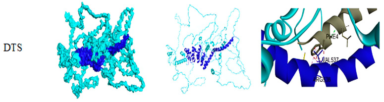
Pathological significance of interaction of Synphilin-1 with mutated alpha-synuclein is well known to have serious consequences in causing the formation of inclusion bodies that are linked to Parkinson's disease (PD). Information extracted so far pointed out that specific mutations, A53T, A30P, and E46K, in alpha-synuclein promote such interactions. However, a detailed structural study of this interaction is pending due to the unavailability of the complete structures of the large protein Synphilin-1 of chain length 919 residues and the mutated alpha-synuclein having all the reported specific mutations so far. In this study, a semi-automatic pipeline-based meta-predictor, AlphaLarge, is created to predict high-fidelity structures of large proteins like Synphilin-1 given the limitations of the existing protocols. AlphaLarge recruits a novel augmented AlphaFold model that uses a divide and conquer based strategy on the foundation of a self-sourced template dataset to choose the best structure model through their standard validations. The structure models were re-validated by a Protein Mediated Interaction Analysis (PMIA) formalism that uses the existing structurally relevant information of these proteins. For the training dataset, the new method, AlphaLarge, performed reasonably better than AlphaFold. Also, the new residue- and domain-based structural details of interactions of resultant best structure models of Synphilin-1 and both wild and mutated alpha-synuclein are extracted using PMIA. This result paves the way for better screening of target specific drugs to control the progression of PD, in particular, and research on any kind of pathophysiology involving large proteins of unknown structures, in general.
Keywords: Lewy bodies; Parkinson’s disease; Synphilin-1 and alpha-synuclein interaction; prediction of large protein structures; protein aggregates; protein–protein interaction.
Conflict of interest statement
The authors declare no conflicts of interest.
Figures


Similar articles
-
alpha-Synuclein is colocalized with 14-3-3 and synphilin-1 in A53T transgenic mice.Acta Neuropathol. 2006 Dec;112(6):681-9. doi: 10.1007/s00401-006-0132-2. Epub 2006 Sep 7. Acta Neuropathol. 2006. PMID: 16957925
-
Synphilin isoforms and the search for a cellular model of lewy body formation in Parkinson's disease.Cell Cycle. 2006 Sep;5(18):2082-6. doi: 10.4161/cc.5.18.3209. Epub 2006 Sep 15. Cell Cycle. 2006. PMID: 16969096 Review.
-
Overexpression of synphilin-1 promotes clearance of soluble and misfolded alpha-synuclein without restoring the motor phenotype in aged A30P transgenic mice.Hum Mol Genet. 2014 Feb 1;23(3):767-81. doi: 10.1093/hmg/ddt467. Epub 2013 Sep 24. Hum Mol Genet. 2014. PMID: 24064336
-
Interaction with synphilin-1 promotes inclusion formation of alpha-synuclein: mechanistic insights and pathological implication.FASEB J. 2010 Jan;24(1):196-205. doi: 10.1096/fj.09-133082. Epub 2009 Sep 17. FASEB J. 2010. PMID: 19762560
-
Synphilin-1 isoforms in Parkinson's disease: regulation by phosphorylation and ubiquitylation.Cell Mol Life Sci. 2008 Jan;65(1):80-8. doi: 10.1007/s00018-007-7343-0. Cell Mol Life Sci. 2008. PMID: 17982729 Free PMC article. Review.
References
-
- Kouli A., Torsney K.M., Kuan W.L. Parkinson’s Disease: Etiology, Neuropathology, and Pathogenesis. In: Stoker T.B., editor. Parkinson’s Disease: Pathogenesis and Clinical Aspects. Codon Publications; Singapore: 2018. - PubMed
MeSH terms
Substances
LinkOut - more resources
Full Text Sources
Medical
Miscellaneous
