. 2024 Dec 16;14(12):1604.
doi: 10.3390/biom14121604.

Dissecting Cytophagalysin: Structural and Biochemical Studies of a Bacterial Pappalysin-Family Metallopeptidase

Affiliations

Dissecting Cytophagalysin: Structural and Biochemical Studies of a Bacterial Pappalysin-Family Metallopeptidase

Eva Estevan-Morió et al. Biomolecules. .

Abstract

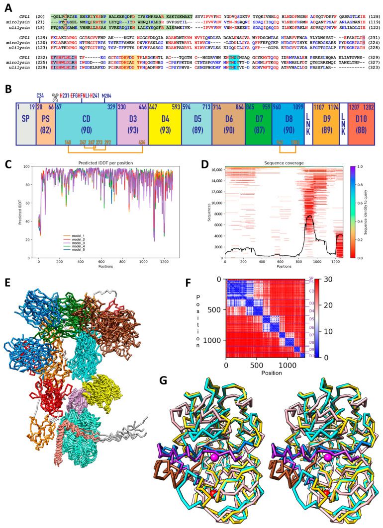
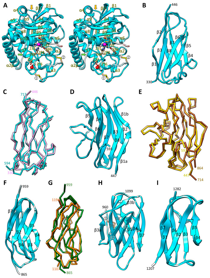
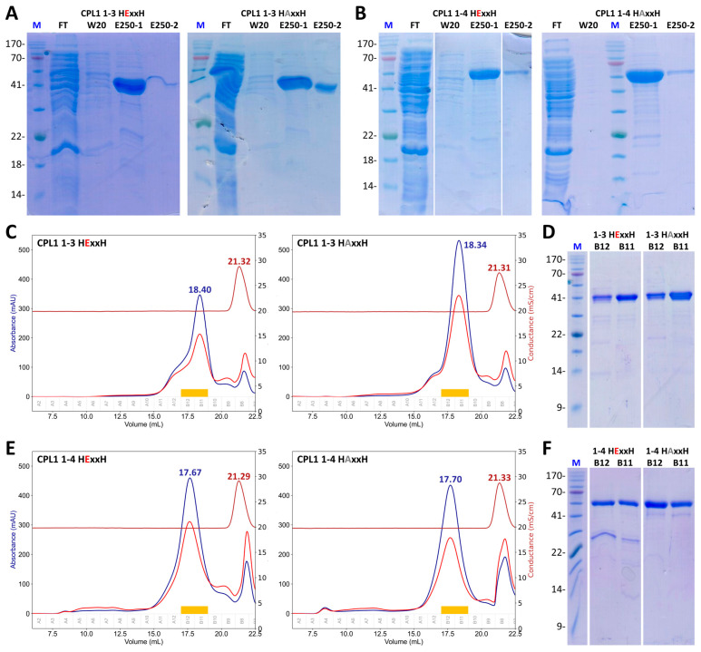
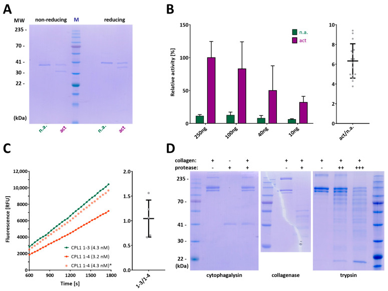
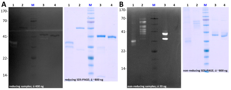
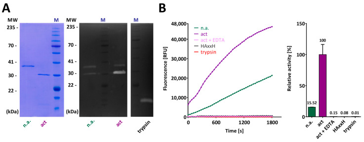
Cytophaga is a genus of Gram-negative bacteria occurring in soil and the gut microbiome. It is closely related to pathogenic Flavobacterium spp. that cause severe diseases in fish. Cytophaga strain L43-1 secretes cytophagalysin (CPL1), a 137 kDa peptidase with reported collagenolytic and gelatinolytic activity. We performed highly-confident structure prediction calculations for CPL1, which identified 11 segments and domains, including a signal peptide for secretion, a prosegment (PS) for latency, a metallopeptidase (MP)-like catalytic domain (CD), and eight immunoglobulin (Ig)-like domains (D3-D10). In addition, two short linkers were found at the D8-D9 and D9-D10 junctions, and the structure would be crosslinked by four disulfide bonds. The CPL1 CD was found closest to ulilysin from Methanosarcina acetivorans, which assigns CPL1 to the lower-pappalysin family within the metzincin clan of MPs. Based on the structure predictions, we aimed to produce constructs spanning the full-length enzyme, as well as PS+CD, PS+CD+D3, and PS+CD+D3+D4. However, we were successful only with the latter three constructs. We could activate recombinant CPL1 by PS removal employing trypsin, and found that both zymogen and mature CPL1 were active in gelatin zymography and against a fluorogenic gelatin variant. This activity was ablated in a mutant, in which the catalytic glutamate described for lower pappalyins and other metzincins was replaced by alanine, and by a broad-spectrum metal chelator. Overall, these results proved that our recombinant CPL1 is a functional active MP, thus supporting the conclusions derived from the structure predictions.

Keywords: functional characterization; metallopeptidase; pappalysin family; protease activation; recombinant protein expression.


Conflict of interest statement

The authors declare no financial or non-financial conflicts of interest with the contents of this article, and the funders had no involvement in the study’s design, data collection, analysis, or interpretation, manuscript preparation, or the decision to publish the findings.

Figures

Figure 1
Figure 1
Biocomputational studies. (A) Sequence alignment of the prosegments (PSs) (green background) and catalytic domains (CDs) of CPL1 (UP Q46348), mirolysin (UP G8ULV1), and ulilysin (UP Q8TL28). Identical or equivalent residues are in red, and those shared by two sequences are in blue. The PS cysteine engaged in zinc-binding in the zymogen—putatively in CPL1—is framed. The extended zinc-binding motif, the residues shaping the common calcium site, and the Met-turn motif are shown over light-blue, orange, and cyan background, respectively. (B) Domain distribution along the chemical sequence predicted by AlphaFold, which foresees a signal peptide for secretion (SP), the PS, the CD, and immunoglobulin-like domains D3 through D10. Each domain is labelled, the respective limiting residues are indicated, and the average predicted local-distance difference test (pLDDT) is shown in parenthesis. In all cases, these values are close to or exceed the high-accuracy cut-off of ~90% [42], and are thus classed as high confidence. The only exception is the PS, whose prediction evinces an average pLDDT that is slightly lower, but still highly reliable for the main chain. Two short linkers (LNKs) would be intercalated between D8 and D9, and between D9 and D10. Predicted disulfide bonds are shown in orange. The cysteine putatively engaged in latency in the zymogen (C24) and the extended zinc-binding motif (H231–H241), as well as the Met-turn methionine (M284) and the maturation cleavage point (A66–E67; scissors) are further pinpointed. (C) pLDDT for each residue of the prediction (positions 1–1282) for each of the five distinct models obtained. (D) Sequence coverage for each residue of the prediction (positions 1–1282) vs. number of sequences. (E) Superposition of the five predicted models without further relaxation/minimization with each domain/segment in the colour of (B). Only PS, CD, D3, D4, and, roughly, D5 appear with similar relative orientations in all models. (F) Analysis of the predicted aligned error, which estimates if domains are correctly positioned relative to one another, for each residue of the prediction (positions 1–1282; model_1). Each segment/domain of (B) gives rise to a marine blue square along the diagonal. Off-diagonal blue values suggest well-predicted interactions between domains. (G) Superposition of the Cα-traces of the experimental structures of promirolysin (PS in sienna, CD in gold) and proulilysin (cyan/dodger blue) in standard orientation [15] onto the prediction of CPL1 (purple/pink). The CPL1 prediction matches proulilysin significantly better. The catalytic zinc (magenta sphere) and the common calcium (red sphere) of proulilysin are further displayed.
Figure 2
Figure 2
Structural analysis of the predicted CPL1 domains. (A) Ribbon-type plot of the CPL1 PS and CD in cross-eye stereo. The secondary structure elements are labelled (α1p, α2p, α1–α9, and β1–β6). The putative cysteine-switch cysteine (C24), zinc-binding residues (H231, H235 and H241), general base/acid glutamate (E232), Met-turn methionine (M284) and tyrosine-switch tyrosine (Y286), calcium-binding residues (D251 and T256), as well as the putative disulfide-bonded cysteines (C247–C273; ① and C267–C292; ②) are shown for their side chains as sticks and numbered. The zinc and calcium cations were modelled based on the proulilysin (PDB 8CDB) and mature ulilysin (PDB 2CKI) structures. The putative maturation site (A66–E67) and the LNR-loop are highlighted by green and orange arrows, respectively. Depiction of the Ig-like domains (D3–D10) showing as ribbon- or Cα-plots (B) D3; (C) D5 (cyan Cα-plot) onto D3 (plum Cα-plot) in the same orientation as in (B); (D) D4; (E) D6 (brown Cα-plot) onto D4 (yellow Cα-plot) in the same orientation as (D); (F) D7; (G) D9 (orange Cα-plot) onto D7 (green Cα-plot) in the same orientation as (F); (H) D8 (disulfide bond C963–C1083; ①) and (I) D10. The β-strands and the N- and C-terminal residues are numbered in all cases.
Figure 3
Figure 3
Recombinant CPL1 expression and purification. (A) Representative SDS-PAGE gels of nickel-affinity purifications of construct CPL1_1-3 (Q20–V444), both in its wild-type (left panel) and E232A-mutant (right panel) variants. Samples representing the flow through (FT), the wash-step with 20 mM imidazole (W20), and the first two elution fractions using 250 mM imidazole (E250-1/2) were analysed under reducing conditions. Lane M depicts the molecular-mass marker. The target protein migrated as a band at its expected molecular weight (~47 kDa). (B) Same as (A) for construct CPL1_1-4 (Q20–T591), which migrates as a ~63 kDa band as expected. (C) Representative calibrated size-exclusion chromatography profiles of the two constructs of (A) using bovine-serum albumin as the calibration standard, and with the conductivity trace shown in dark red (peak at ~21.3 mL), and (D) SDS-PAGE analyses of peak fractions B11 and B12 shown in (C) as orange bands. A retention volume of ~18.4 mL corresponds to an apparent molecular mass of ~41 kDa, which is consistent with the theoretic value (~47 kDa). (E,F) Same as (C,D) for the two constructs of (B). A retention volume of ~17.7 mL corresponds to an apparent molecular weight of ~61 kDa, which is consistent with the theoretic value (~63 kDa). SDS-PAGE gels were cropped for clarity. For full gel images, please refer to Extended Data Figures S7–S10 in the supplement.
Figure 4
Figure 4
Trypsin-mediated activation and activity of CPL1 constructs. (A) SDS-PAGE analysis under non-reducing and reducing conditions, which shows that the trypsin-activated protease sample (act) shows a band ~7 kDa smaller than the non-activated sample (n.a.), which corresponds to the excision of the zymogenic N-terminal prosegment. (B) (Left) Average and standard deviation of relative activity of different amounts of activated wild-type CPL1_1-3 against the fluorogenic substrate DQ Gelatin (2 μg) compared to the non-activated zymogen. (Right) Ratio of activities between both protein variants (n = 16). (C) (Left) Fluorescence resulting from the turnover of DQ Gelatin by activated wild-type CPL1_1-3 and CPL1_1-4. The values shown in salmon for the latter are recalculated from the recorded curve at 3.2 nM and correspond to the same concentration as those for CPL1_1-3, and are therefore marked with an asterisk (*). (Right) Normalized molarity values for the two constructs (n = 5), which reveal equivalent activity. (D) SDS-PAGE analysis of the incubation of human type-I atelocollagen (10 μg) with 2 μg of wild-type CPL1_1-3 (left), 0.5 μg of Clostridium histolyticum collagenase (centre), and 0.1 μg (++) or 1 μg (+++) of bovine trypsin (right). The + and − signs indicate the presence or absence of collagen and the respective protease (cytophagalysin, collagenase, or trypsin, as labeled below the gels), with increasing + signs denoting higher protease concentrations. SDS-PAGE gels were cropped for clarity. For full gel images, please refer to Extended Data Figures S11 and S12 in the supplementary materials.
Figure 5
Figure 5
CPL1 activity in gelatin zymography. (A) Representative gelatin zymograms (left) and SDS-PAGE analysis (right) under reducing conditions of wild-type variants CPL1_1-2 (lane 1), CPL1_1-4 (lane 2), and CPL1_1-3 (lane 3), which evince only minute activity due to the unfolding of the protein variants caused by the reducing conditions, as well as of inactive CPL1_1-3 E232A-mutant (lane 4). (B) Same as (A) under non-reducing conditions, which locally preserves the integrity of the recombinant proteins, thereby aiding in-gel refolding and consequently revealing significant activity for wild-type CPL1_1-2 (lane 1), CPL1_1-4 (lane 2), and CPL1_1-3 (lane 3), but not for the mutationally inactivated CPL1_1-3 variant (lane 4). SDS-PAGE gels and zymograms were cropped for clarity. For full gel and zymogram images, please refer to Extended Data Figures S13 and S14 in the supplement.
Figure 6
Figure 6
Inhibition of activity of CPL1. (A) (Left) Complete activation of CPL1_1-3 (lane act) by trypsin in SDS-PAGE, as shown by the absence of the zymogen band that is found in lane n.a. In zymography (Centre), both samples exhibited activity for both activated and non-activated CPL1, with significantly increased activity observed in the activated sample. Importantly, no trypsin activity was detected in the CPL1 samples. For reference, trypsin activity (Right) at an apparent molecular weight of ~18 kDa is shown. In zymography however, both samples demonstrated activity for both activated and non-activated CPL1 (Centre), with activity enriched in the activated sample. Note, no trypsin activity was observed in CPL1 samples, and trypsin activity is shown at an apparent molecular weight of ~18 kDa (Right). (B) The activity of CPL1_1-3 against fluorogenic DQ Gelatin is efficiently inhibited by EDTA as expected for a metallopeptidase, yielding only residual values that are comparable to those of the E232A-mutant and trypsin, which does not cleave this substrate. Note that the CPL1_1-3 zymogen still has a residual activity of ~15% of the active form. SDS-PAGE gels and zymograms were cropped for clarity. For full gel and zymogram images, please refer to Extended Data Figure S15 in the supplementary materials.

Similar articles

References

    1. Klein T., Eckhard U., Dufour A., Solis N., Overall C.M. Proteolytic Cleavage-Mechanisms, Function, and “Omic” Approaches for a Near-Ubiquitous Posttranslational Modification. Chem. Rev. 2018;118:1137–1168. doi: 10.1021/acs.chemrev.7b00120. - DOI - PubMed
    1. Fushimi N., Ee C.E., Nakajima T., Ichishima E. Aspzincin, a Family of Metalloendopeptidases with a New Zinc-Binding Motif. Identification of New Zinc-Binding Sites (His128, His132, and Asp164) and Three Catalytically Crucial Residues (Glu129, Asp143, and Tyr106) of Deuterolysin from Aspergillus Oryzae by Site-Directed Mutagenesis. J. Biol. Chem. 1999;274:24195–24201. doi: 10.1074/jbc.274.34.24195. - DOI - PubMed
    1. Hooper N.M. Families of Zinc Metalloproteases. FEBS Lett. 1994;354:1–6. doi: 10.1016/0014-5793(94)01079-X. - DOI - PubMed
    1. Bode W., Gomis-Rüth F.X., Stöcker W. Astacins, Serralysins, Snake Venom and Matrix Metalloproteinases Exhibit Identical Zinc-Binding Environments (HEXXHXXGXXH and Met-Turn) and Topologies and Should Be Grouped into a Common Family, the ‘metzincins’. FEBS Lett. 1993;331:134–140. doi: 10.1016/0014-5793(93)80312-I. - DOI - PubMed
    1. Schneider J.S., Glickman M.S. Function of Site-2 Proteases in Bacteria and Bacterial Pathogens. Biochim. Biophys. Acta. 2013;1828:2808–2814. doi: 10.1016/j.bbamem.2013.04.019. - DOI - PMC - PubMed

MeSH terms

LinkOut - more resources