. 2024 Dec 20;14(12):1642.
doi: 10.3390/biom14121642.

Gramicidin A in Asymmetric Lipid Membranes

Affiliations

Gramicidin A in Asymmetric Lipid Membranes

Oleg V Kondrashov et al. Biomolecules. .

Abstract

Gramicidin A is a natural antimicrobial peptide produced by Bacillus brevis. Its transmembrane dimer is a cation-selective ion channel. The channel is characterized by the average lifetime of the conducting state and the monomer-dimer equilibrium constant. Dimer formation is accompanied by deformations of the membrane. We theoretically studied how the asymmetry in lipid membrane monolayers influences the formation of the gramicidin A channel. We calculated how the asymmetry in the spontaneous curvature and/or lateral tension of lipid monolayers changes the channel lifetime and shifts the equilibrium constant of the dimerization/dissociation process. For the asymmetry expected to arise in plasma membranes of mammalian cells upon the addition of gramicidin A or its derivatives to the cell exterior, our model predicts a manifold increase in the average lifetime and equilibrium constant.

Keywords: asymmetric lipid membrane; channel lifetime; equilibrium constant; gramicidin A; intrinsic curvature; lateral tension; lipid–protein interaction; membrane biophysics; theory of elasticity.


Conflict of interest statement

The authors declare no conflict of interest. The funders had no role in the design of the study; in the collection, analyses, or interpretation of data; in the writing of the manuscript; or in the decision to publish the results.

Figures

Figure 1
Figure 1
Configurations of two gA molecules located in opposing monolayers of the membrane: two monomers (left); conducting dimer (right); and coaxial pair (middle, top). The elastic energy of the membrane in these configurations is shown schematically. The states of two monomers and the conducting dimer are stable and metastable, respectively. These two configurations are in equilibrium with each other. The coaxial pair corresponds to the top of the energy barrier of the dimerization/dissociation process. The energy barrier of dimerization is the difference in the energies of the coaxial pair and two monomers; the energy barrier of dissociation is the difference in the energies of the coaxial pair and dimer. The only ion-conducting configuration is the dimer. Ionic conductance is harmful to cells as it leads to homeostasis violation.
Figure 2
Figure 2
Dependences of logarithms of normalized dimer lifetime τ0/τ00 and normalized equilibrium constant K0/K00 at almost zero lateral tensions, σu = σl ≈ 0 (corresponding to plasma membranes of cells or deflated GUVs) on spontaneous curvatures of the outer (Ju) and inner (Jl) monolayers. Larger K0 corresponds to larger equilibrium number of dimers and higher integral conductance of the membrane.
Figure 3
Figure 3
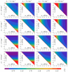
Dependence of normalized gA dimer lifetime τ/τ0 on lateral tensions in the outer (σu) and inner (σl) monolayers for different values of spontaneous curvature of the outer (Ju) and inner (Jl) monolayers. The values of τ0 were obtained as the limit of τ when (σu, σl) → (0, 0), corresponding to plasma membranes of cells or deflated GUVs. In white triangles in left-lower corners of the plots, σu + σl < 0, and the membrane is mechanically unstable. Thus, only right-upper halves of the plots are displayed.
Figure 4
Figure 4
Dependence of normalized gA dimer–monomer equilibrium constant K/K0 on lateral tensions in the outer (σu) and inner (σl) monolayers for different values of spontaneous curvature of the outer (Ju) and inner (Jl) monolayers. The values of K0 were obtained as the limit of K when (σu, σl) → (0, 0), corresponding to plasma membranes of cells or deflated GUVs. In white triangles in left-lower corners of the plots, σu + σl < 0, and the membrane is mechanically unstable. Thus, only right-upper halves of the plots are displayed.

References

    1. Hladky S.B., Haydon D.A. Ion transfer across lipid membranes in the presence of gramicidin A: I. Studies of the unit conductance channel. Biochim. Biophys. Acta. 1972;274:294–312. doi: 10.1016/0005-2736(72)90178-2. - DOI - PubMed
    1. Bamberg E., Apell H.J., Alpes H. Structure of the gramicidin A channel: Discrimination between the πL, D and the β helix by electrical measurements with lipid bilayer membranes. Proc. Natl. Acad. Sci. USA. 1977;74:2402–2406. doi: 10.1073/pnas.74.6.2402. - DOI - PMC - PubMed
    1. Arseniev A.S., Barsukov I.L., Bystrov V.F., Lomize A.L., Ovchinnikov Y.A. 1H-NMR study of gramicidin A transmembrane ion channel: Head-to-head right-handed, single-stranded helices. FEBS Lett. 1985;186:168–174. doi: 10.1016/0014-5793(85)80702-X. - DOI - PubMed
    1. Andersen O.S., Apell H.J., Bamberg E., Busath D.D., Koeppe R.E., Sigworth F.J., Szabo G., Urry D.W., Woolley A. Gramicidin channel controversy—The structure in a lipid environment. Nat. Struct. Mol. Biol. 1999;6:609. doi: 10.1038/10648. - DOI - PubMed
    1. Bamberg E., Läuger P. Channel formation kinetics of gramicidin A in lipid bilayer membranes. J. Membr. Biol. 1973;11:177–194. doi: 10.1007/BF01869820. - DOI - PubMed

MeSH terms

LinkOut - more resources