. 2024 Nov 26;14(12):1192.
doi: 10.3390/brainsci14121192.

Erlotinib Improves the Response of Glioblastoma Cells Resistant to Photodynamic Therapy

Affiliations

Erlotinib Improves the Response of Glioblastoma Cells Resistant to Photodynamic Therapy

Karen Olthoff et al. Brain Sci. .

Abstract

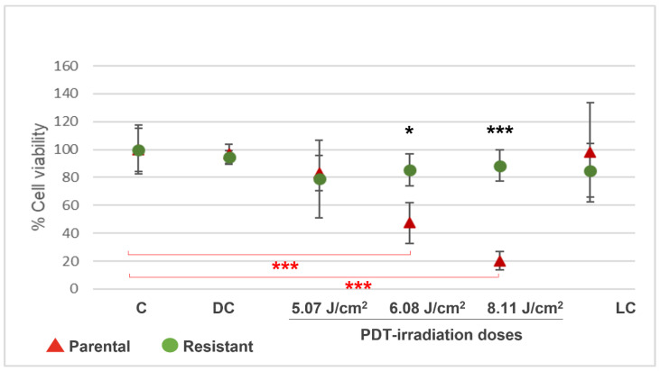
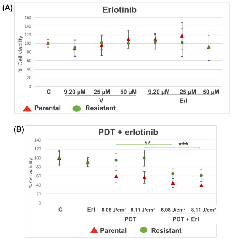
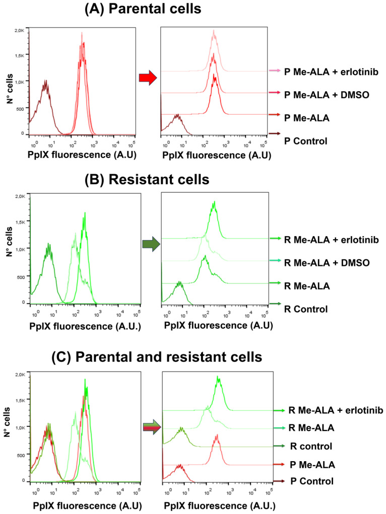
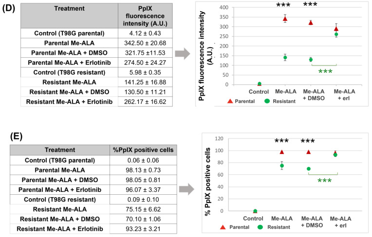
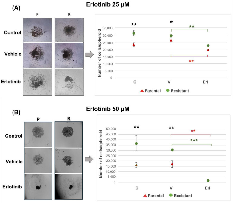
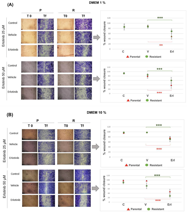
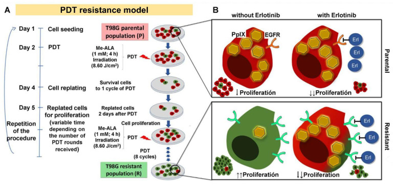
Background: Glioblastoma (GBM) is the most common and deadly type of brain cancer in adults. Dysregulation of receptor tyrosine kinase pathways, such as the epidermal growth factor receptor (EGFR), contributes to therapeutic resistance. Drugs that inhibit tyrosine kinase activity and monoclonal antibodies against EGFR are strategies used in clinical trials. Photodynamic therapy (PDT) is a tumor treatment that involves the administration of a photosensitizing drug, followed by its activation with visible light, which causes cell death due to oxidative stress. Although PDT helps prolong median survival in patients with GBM, complete remission has not been achieved. Populations of GBM cells have been obtained from the T98G line resistant to PDT with methyl-5-aminolevulinic acid (Me-ALA) for characterization, comparing them with the original parental population. Objective: The objective of this work was to evaluate the general response of T98G GBM cells resistant to PDT when EGFR activity is inhibited with the drug erlotinib. Methods and Results: It has been observed that the administration of the EGFR inhibitor drug in combination with PDT reduced viability (MTT) in resistant populations compared to PDT alone. Furthermore, the PpIX content (flow cytometry) was increased in the resistant population when cells were incubated with Me-ALA and erlotinib. Erlotinib prevented cell proliferation of parental and resistant spheroids. Wound closure was reduced in both parental and PDT-resistant populations. Conclusions: Our results indicate that EGFR activation would be relevant in the resistance of GBM cells to PDT.

Keywords: EGFR; erlotinib; glioblastoma; photodynamic therapy; resistance.


Conflict of interest statement

The authors declare no conflicts of interest.

Figures

Figure 1
Figure 1
Percentages of cell viability by MTT after PDT in T98G parental and PDT-resistant populations. Parental and resistant T98G cells were incubated with Me-ALA for 4 h and irradiated at 5.07, 6.08, and 8.11 J/cm2. Resistant populations had high viability at irradiation doses lethal to the parental populations. C = control (without Me-ALA, without light); DC = drug control (with Me-ALA, without light); LC = 8.11 J/cm2 light control (without Me-ALA, with light, 8.11 J/cm2); photodynamic therapy (PDT)-treated (Me-ALA + 5.07 J/cm2; Me-ALA + 6.08 J/cm2; Me-ALA + 8.11 J/cm2). * p < 0.05; *** p = 0.001 (resistant respect to parental cells, black asterisks), *** p ≤ 0.001 (resistant with PDT respect to resistant C, red asterisks). Results are reported as the mean ± standard error of mean (SEM). The experiment was performed 3 times. Results of a representative experiment are shown.
Figure 2
Figure 2
(A) Percentages of cell viability by MTT in parental and resistant T98G cells incubated with erlotinib. Parental and resistant T98G cells were incubated with erlotinib (Erl) for 24 h at doses of 9.2, 25, and 50 µM. Erlotinib and its vehicle DMSO (V) did not affect the cell viability (at 24 h) of parental and resistant T98G populations. (B) Cell viability by MTT after PDT and erlotinib combination in parental and resistant T98G cells. Cells were incubated with Me-ALA (1 mM) and erlotinib (50 uM) for 4 h and then irradiated at 6.08 and 8.11 J/cm2. After 24 h, cell viability was measured by MTT. PDT in combination with erlotinib reduced viability in resistant populations compared to PDT alone. Photodynamic therapy (PDT)-treated (Me-ALA + 6.08 J/cm2; Me-ALA + 8.11 J/cm2). ** p < 0.01; *** p = 0.001 (resistant populations, green asterisks). Results are reported as the mean ± standard error of mean (SEM). The experiment was performed 3 times. Results of a representative experiment are shown.
Figure 3
Figure 3
Flow cytometry of PpIX content after Me-ALA and erlotinib incubation. Parental (A) and resistant cells (B) were incubated with Me-ALA (1 mM) and erlotinib (50 µM) for 4 h. PpIX fluorescence was measured by flow cytometry. Erlotinib increased the content of PpIX in the resistant population (BE). C = control (without Me-ALA, without erlotinib); Erl = erlotinib; A.U. = arbitrary units. *** p < 0.001 (resistant respect to parental cells, black asterisks), *** p < 0.001 (resistant cells, green asterisks). Results are reported as the mean ± SEM. The experiment was performed 3 times. Results of a representative experiment are shown.
Figure 3
Figure 3
Flow cytometry of PpIX content after Me-ALA and erlotinib incubation. Parental (A) and resistant cells (B) were incubated with Me-ALA (1 mM) and erlotinib (50 µM) for 4 h. PpIX fluorescence was measured by flow cytometry. Erlotinib increased the content of PpIX in the resistant population (BE). C = control (without Me-ALA, without erlotinib); Erl = erlotinib; A.U. = arbitrary units. *** p < 0.001 (resistant respect to parental cells, black asterisks), *** p < 0.001 (resistant cells, green asterisks). Results are reported as the mean ± SEM. The experiment was performed 3 times. Results of a representative experiment are shown.
Figure 4
Figure 4
Parental and resistant T98G spheroids incubated with erlotinib. Photographs and cell number count per spheroid 3 days after seeding 2000 cells per well in U-bottom plates with agarose. Cells were seeded and incubated with erlotinib (Erl) at the dose of 25 µM (A) and 50 µM (B) for 3 days. Erlotinib inhibited the proliferation of parental and resistant T98G 3D cultures. C = control, V = vehicle, Erl = erlotinib. * p < 0.01; ** p < 0.01 (resistant respect to parental cells, black asterisks); ** p < 0.01 (parental cells, green asterisks; resistant cells, red asterisks) and *** p = 0.001 (parental cells, green asterisks). Results are reported as the mean ± SEM. The experiment was performed 3 times. Results of a representative experiment are shown.
Figure 5
Figure 5
Wound closure in parental and resistant T98G populations incubated with erlotinib. Immediately after opening the wounds, cells were incubated with erlotinib (Erl) at the dose of 25 µM in DMEM 1% (A) and DMEM 10% (B) and at the dose of 50 µM in DMEM 1% (A) and DMEM 10% (B). Photographs of the wounds were taken at day 0 and at the final time. A quantification of the percentage of wound closure was performed using ImageJ 1.53t. Erlotinib reduced wound closure similarly in parental and resistant T98G, both when 1% DMEM was used and when 10% DMEM was used. C = control, V = vehicle, Erl = erlotinib. ** p < 0.01, *** p < 0.001 (parental cells, green asterisks; resistant cells, red asterisks). Results are reported as the mean ± SEM. The experiment was performed 3 times. Photographs and results of a representative experiment are shown.
Figure 6
Figure 6
Protocol for obtaining PDT-resistant T98G cells and the effect of erlotinib. (A) Timeline: initial population of T98G cells (parental) is treated with PDT (Me-ALA); the surviving cells are amplified forming a population of resistant cells to one cycle of PDT. The process is repeated for 8 cycles (a selected population resistant to PDT is formed). (B) PDT-resistant T98G cells overexpress EGFR mRNA, accumulate less PpIX and have increased 3D proliferation with respect to parental cells [14]. Treatment with the EGFR inhibitor erlotinib (erl) causes a decrease in 3D cell proliferation (parental and resistant populations). Additionally, when treated with erlotinib, resistant cells regain the ability to accumulate PpIX becoming sensitive to PDT.

Similar articles

References

    1. Grochans S., Cybulska A.M., Siminska D., Korbecki J., Kojder K., Chlubek D., Baranowska-Bosiacka I. Epidemiology of Glioblastoma Multiforme–Literature Review. Cancers. 2022;14:2412. doi: 10.3390/cancers14102412. - DOI - PMC - PubMed
    1. Wu W., Klockow J.L., Zhang M., Lafortune F., Chang E., Jin L., Wu Y., Daldrup-Link H.E. Glioblastoma multiforme (GBM): An overview of current therapies and mechanisms of resistance. Pharmacol. Res. 2021;171:105780. doi: 10.1016/j.phrs.2021.105780. - DOI - PMC - PubMed
    1. Schupper A.J., Rao M., Mohammadi N., Baron R., Lee J.Y.K., Acerbi F., Hadjipanayis C.G. Fluorescence-Guided Surgery: A Review on Timing and Use in Brain Tumor Surgery. Front. Neurol. 2021;12:682151. doi: 10.3389/fneur.2021.682151. - DOI - PMC - PubMed
    1. Ibarra L.E., Vilchez M.L., Caverzán M.D., Milla Sanabria L.N. Understanding the glioblastoma tumor biology to optimize photodynamic therapy: From molecular to cellular events. J. Neurosci. Res. 2021;99:1024–1047. doi: 10.1002/jnr.24776. - DOI - PubMed
    1. Kiesel B., Wadiura L.I., Mischkulnig M., Makolli J., Sperl V., Borkovec M., Freund J., Lang A., Millesi M., Berghoff A.S., et al. Efficacy, Outcome, and Safety of Elderly Patients with Glioblastoma in the 5-ALA Era: Single Center Experience of More Than 10 Years. Cancers. 2021;13:6119. doi: 10.3390/cancers13236119. - DOI - PMC - PubMed

LinkOut - more resources