. 2024 Nov 24;13(12):1127.
doi: 10.3390/antibiotics13121127.

Cloning and Functional Analysis of Skin Host Defense Peptides from Yakushima Tago's Brown Frog (Rana tagoi yakushimensis) and Development of Serum Endotoxin Detection System

Affiliations

Cloning and Functional Analysis of Skin Host Defense Peptides from Yakushima Tago's Brown Frog (Rana tagoi yakushimensis) and Development of Serum Endotoxin Detection System

Taichi Aono et al. Antibiotics (Basel). .

Abstract

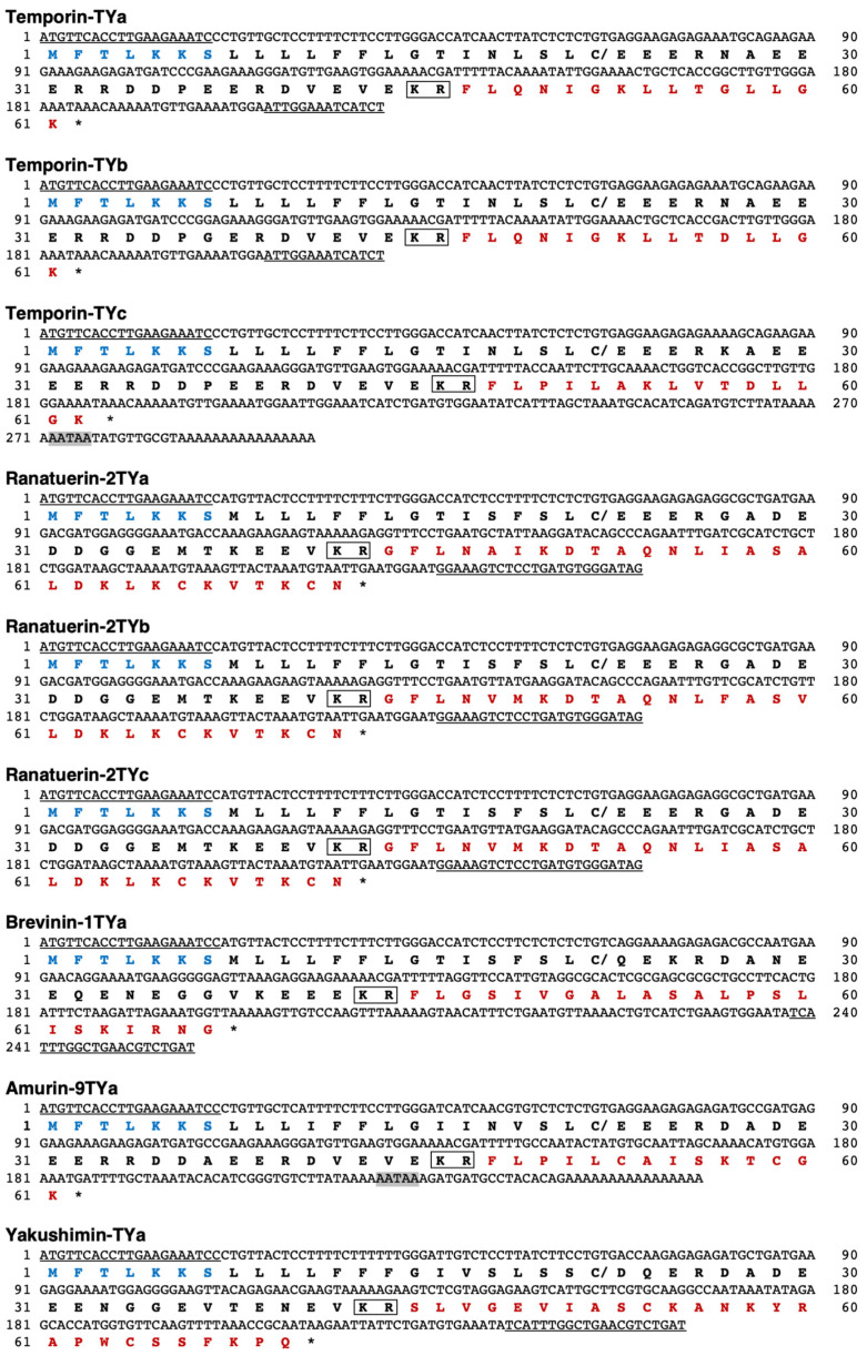
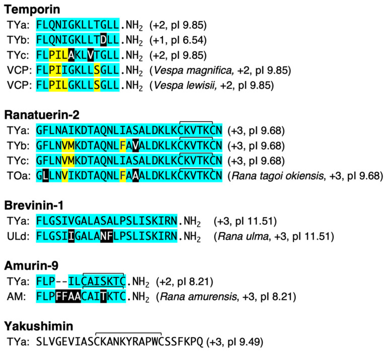
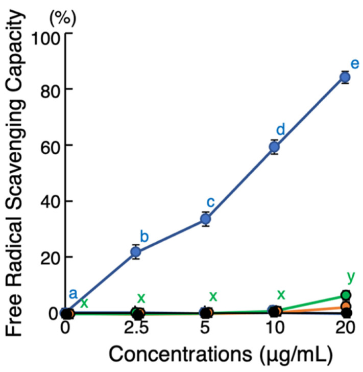
Background/Objective: Amphibian skin is a valuable source of host defense peptides (HDPs). This study aimed to identify HDPs with novel amino acid sequences from the skin of Rana tagoi yakushimensis and analyze their functions. Methods: cDNAs encoding HDP precursors were cloned and sequenced using RT-PCR and 3'-RACE. The novel HDPs were synthesized to evaluate their antimicrobial activity, antioxidant activity, and cytotoxicity. Antimicrobial activity was evaluated by way of broth microdilution and endotoxin- and β-glucan-binding capacity using an enzyme-linked endotoxin binding assay (ELEBA) and a modified ELEBA, respectively. Results: Nine cDNAs encoding precursors for various HDP families, including temporin, ranatuerin-2, brevinin-1, amurin-9, and a novel yakushimin peptide, were identified. Brevinin-1TYa exhibited antibacterial activity against Staphylococcus aureus, and brevinin-1TYa and amurin-9TYa induced morphological changes in Escherichia coli and S. aureus. Yakushimin-TYa, amurin-9TYa, and brevinin-1TYa showed concentration-dependent antibacterial effects against the plant pathogens Xanthomonas oryzae pv. oryzae and Clavibacter michiganensis subsp. michiganensis. Amurin-9TYa demonstrated strong binding affinity to lipopolysaccharide, lipoteichoic acid, and β-glucan, exhibited antioxidant activity, and lacked cytotoxicity, making it a promising therapeutic candidate. Moreover, brevinin-1TYa showed strong cytotoxicity, whereas yakushimin-TYa exhibited weak cytotoxicity. Conclusions: These findings highlight the potential of these peptides, particularly amurin-9TYa, for future applications as antimicrobial and therapeutic agents.

Keywords: ELEBA; ELGBA; Rana tagoi yakushimensis; amurin-9; anti-plant pathogenic activity; antioxidant activity; brevinin-1; frog skin; host defense peptides; yakushimin.


Conflict of interest statement

The authors declare no conflicts of interest.

Figures

Figure 1
Figure 1
Nucleotide and deduced amino acid sequences of HDP precursor cDNAs that were cloned from R. t. yakushimensis skin; in the nucleotide sequences, the forward and reverse primer-derived sequences are underlined. The putative polyadenylation signals are indicated with gray In the amino acid sequences, the forward primer-derived and HDP sequences are indicated with blue and red letters, respectively. Stop codons are marked with asterisks (*), and cleavage sites for signal peptidase and processing enzymes are marked with slashes (/) and boxes, respectively.
Figure 2
Figure 2
Comparison and schematic alignment of the amino acid sequences of the deduced mature temporin, ranatuerin-2, brevinin-1, amurin-9, and yakushimin peptides, as well as other peptides that exhibited the highest sequence similarities in a BLAST search. Residues that are conserved among each peptide are marked by the same color. The reference sequences of each peptide family are provided at the top of each block; generally, these are the originally isolated sequences. Gaps (-) were introduced to maximize sequence identities. Species names (except for R. t. yakushimensis peptides) and the values of the deduced net positive charge and pI are shown in parentheses.
Figure 3
Figure 3
(A) Effects of various concentrations of yakushimin-TYa (blue), amurin-9TYa (green), and brevinin-1TYa (orange) on the growth of Escherichia coli and Staphylococcus aureus. Bacterial cells were incubated with twofold serially diluted peptides for 20 h at 37 °C. Points and vertical bars represent the mean ± standard error of the mean (SE); n = 4. Values with the same superscripts are not significantly different (p ≥ 0.05). (B) Scanning electron microscopy (SEM) analysis of E. coli and S. aureus cells treated with yakushimin-TYa, amurin-9TYa, or brevinin-1TYa. Bacterial cultures in the mid-log phase were incubated with the various peptides (128 μg/mL) for 1 h at 25 °C and examined by SEM. Abnormally shaped cells indicating cell destruction (arrows) are visible in amurin-9TYa-treated E. coli and S. aureus, and brevinin-1TYa-treated S. aureus, compared to the myoglobin-treated controls. Scale bar = 1 μm.
Figure 4
Figure 4
Effects of various concentrations of yakushimin-TYa (blue), amurin-9TYa (green), and brevinin-1TYa (orange) on the growth of plant pathogenic Gram-negative bacteria Xanthomonas oryzae pv. oryzae and Gram-positive bacteria Clavibacter michiganensis subsp. michiganensis. Cells of each bacterial strain were incubated with serially diluted yakushimin-TYa, amurin-9TYa, or brevinin-1TYa peptides for 24 h at 28 °C. Points and vertical bars represent the mean ± SE (n = 4). In the panels, values with the same superscript are not significantly different (p ≥ 0.05).
Figure 5
Figure 5
(A) Evaluation of the binding abilities of yakushimin-TYa (blue), amurin-9TYa (green), and brevinin-1TYa (orange) to the bacterial endotoxins lipopolysaccharide (LPS) and lipoteichoic acid (LTA) using an enzyme-linked endotoxin binding assay (ELEBA). Twofold serially diluted peptides were coated onto 96-well Immobilizer plates and incubated with biotin-LPS or biotin-LTA for 4 h at 25 °C. Detection was performed using streptavidin-conjugated horseradish peroxidase (SA-HRP) and TMB substrate, and absorbance was measured at 450 nm. (B) Assessment of the competitive binding of LPS (dark green) and LTA (light green) to amurin-9TYa and verification in serum. Amurin-9TYa (40 µg/mL) was incubated on Immobilizer plates at 4 °C for 16 h. Biotin-LPS was mixed with LPS in PBS or FBS (500 ng/mL), and biotin-LTA was mixed with non-labeled LTA, both in blocking reagents. For the competition assays with LTA in FBS, a chloroform extraction was performed. The mixture was stirred and centrifuged, and the supernatant was collected, vaporized, and redissolved in endotoxin-free water for the competition experiment. Columns and vertical bars represent the mean ± SE (n = 4). Bovine serum albumin (BSA; white) was used as a control. Values with the same letters are not significantly different (p ≥ 0.05).
Figure 6
Figure 6
Evaluation of the binding abilities of yakushimin-TYa (blue), amurin-9TYa (green), and brevinin-1TYa (orange) to the fungal cell surface substance β-glucan using a modified ELEBA method, the enzyme-linked β-glucan-binding assay (ELGBA). This method replaces the biotin-labeled endotoxins in the ELEBA method with biotin-labeled β-glucan. The other difference is that the reaction with SA-HRP was extended to 40 min and that with the TMB chromogenic solution to 25 min. Other reaction conditions and details of the preparation of biotin-labeled β-glucan are described in the Section 4. Columns and vertical bars represent the mean ± SE (n = 4). BSA (white) was used as a control. In each panel, values with the same letters are not significantly different (p ≥ 0.05).
Figure 7
Figure 7
Evaluation of antioxidant activities of yakushimin-TYa (blue), amurin-9TYa (green), and brevinin-1TYa (orange). Twofold serially diluted peptide solutions were incubated with ABTS+ in a U-bottom 96-well microplate at 25 °C for 10 min. After incubation, the A734 values were measured and the inhibition percentage of ABTS was calculated. Points and vertical bars represent the mean ± SE (n = 4). Values with the same letters are not significantly different (p ≥ 0.05). Neither brevinin-1TYa nor granuliberin-SSa (black; negative control) [26] exhibited any antioxidant activity.
Figure 8
Figure 8
(A) Evaluation of cytotoxic effects of yakushimin-TYa (blue), amurin-9TYa (green), and brevinin-1TYa (orange) in eukaryotic cells. Media containing 5 × 10³ COS7 cells or 1 × 10⁴ HUVECs in 100 μL aliquots were incubated with twofold serially diluted peptides for 2 h at 37 °C. Cell proliferation was determined using an MTT assay, and the survival rates were expressed relative to the control (0 dose). Columns and vertical bars represent the mean ± SE (n = 4). Values with the same superscripts are not significantly different (p ≥ 0.05). (B) SEM analysis of COS7 cells and HUVECs treated with yakushimin-TYa (128 μg/mL), amurin-9TYa (64 μg/mL), brevinin-1TYa (128 μg/mL), or myoglobin (128 μg/mL, negative control) for 1 h at 25 °C. Abnormally shaped cells, such as those showing cell shrinkage, surface destruction (arrows), or nucleus destruction (arrowhead), are visible in all panels except in the amurin-9TYa-treated HUVECs, compared to the myoglobin-treated cells. Scale bar = 20 μm.

Similar articles

References

    1. O’Neill J. The Review on Antimicrobial Resistance. Wellcome Trust and HM Government; London, UK: 2016. [(accessed on 8 November 2024)]. Tackling drug-resistant infections globally: Final report and recommendations; pp. 1–84. Available online: https://amr-review.org/sites/default/files/160518_Final%20paper_with%20c....
    1. Popova K.B., Valsamatzi-Panagiotou A., Penchovsky R. New drug discovery strategies for targeting drug-resistant bacteria. Environ. Chem. Lett. 2021;19:1995–2004. doi: 10.1007/s10311-021-01181-3. - DOI
    1. Li Y., Qi R., Wang X., Yuan H. Recent strategies to develop conjugated polymers for detection and therapeutics. Polymers. 2023;5:3570. doi: 10.3390/polym15173570. - DOI - PMC - PubMed
    1. Singh V., Chibale K. Strategies to combat multi-drug resistance in Tuberculosis. Acc. Chem. Res. 2021;54:2361–2376. doi: 10.1021/acs.accounts.0c00878. - DOI - PMC - PubMed
    1. Ting D.S.J., Beuerman R.W., Dua H.S., Lakshminarayanan R., Mohammed I. Strategies in translating the therapeutic potentials of host defense peptides. Front. Immunol. 2020;11:983. doi: 10.3389/fimmu.2020.00983. - DOI - PMC - PubMed

Grants and funding

LinkOut - more resources