Case Reports
. 2024 Nov 23;15(12):1503.
doi: 10.3390/genes15121503.

Rescue of Aberrant Splicing Caused by a Novel Complex Deep-intronic ABCA4 Allele

Affiliations
Case Reports

Rescue of Aberrant Splicing Caused by a Novel Complex Deep-intronic ABCA4 Allele

Jordi Maggi et al. Genes (Basel). .

Abstract

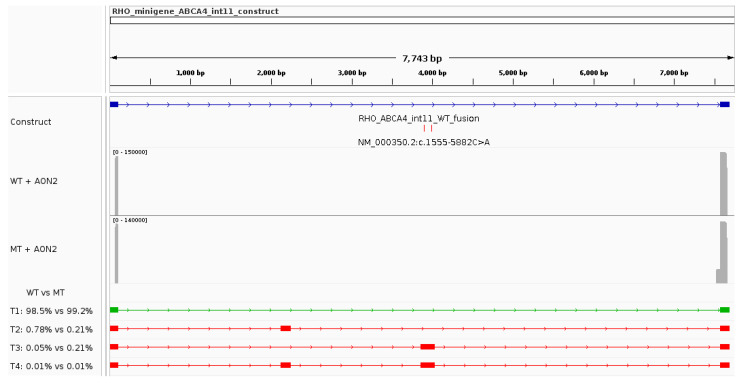
Background/Objectives: Stargardt disease (STGD1) is an autosomal recessive disorder caused by pathogenic variants in ABCA4 that affects the retina and is characterised by progressive central vision loss. The onset of disease manifestations varies from childhood to early adulthood. Methods: Whole exome (WES), whole gene, and whole genome sequencing (WGS) were performed for a patient with STGD1. Results: WES revealed a heterozygous pathogenic missense variant in ABCA4, but no second pathogenic variant was found. ABCA4 whole-gene sequencing, subsequent WGS, and segregation analysis identified a complex deep-intronic allele (NM_000350.2(ABCA4):c.[1555-5882C>A;1555-5784C>G]) in trans to the missense variant. Minigene assays combined with nanopore sequencing were performed to characterise this deep-intronic complex allele in more detail. Surprisingly, the reference minigene revealed the existence of two pseudoexons in intron 11 of the ABCA4 gene that are included in low-abundance (<1%) transcripts. Both pseudoexons could be confirmed in cDNA derived from wildtype retinal organoids. Despite mild splicing predictions, the variant minigene revealed that the complex deep-intronic allele substantially increased the abundance of transcripts that included the pseudoexon overlapping with the variants. Two antisense oligonucleotides (AONs) were designed to rescue the aberrant splicing events. Both AONs increased the proportion of correctly spliced transcripts, and one of them rescued correct splicing to reference levels. Conclusions: Minigene assays combined with nanopore sequencing proved instrumental in identifying low-abundance transcripts including pseudoexons from wildtype ABCA4 intron 11, one of which was substantially increased by the complex allele.

Keywords: ABCA4; Stargardt disease; antisense oligonucleotide; complex allele; deep-intronic variant; minigene; pseudoexon; rescue; retinal organoid; splicing.


Conflict of interest statement

The authors declare no conflicts of interest.

Figures

Figure 1
Figure 1
Fundus autofluorescence (FAF; top left), multifocal electroretinogram (MF-ERG; bottom left), and optical coherence tomography (OCT; right) findings from the index patient at 10 and 16 years of age. Worsening of the FAF and OCT structural findings was confirmed, however, the functional MF-ERG findings were approximately stable (although not directly comparable due to a change of monitor between examinations and likely eccentric fixation).
Figure 2
Figure 2
Visual representation of splicing prediction algorithms from the deep-intronic complex allele in ABCA4. The figure was created from two screenshots of the Alamut Visual Plus software v.1.6.1. (A) The panel shows the genomic region surrounding the complex allele NM_000350.2:c.[1555-5882C>A;1555-5784C>G] (reference sequence above the variant sequence), with the respective splice site predictions computed by the algorithms included in Alamut Visual Plus (SpliceSiteFinder-like, MaxEntScan, NNSPLICE, and GeneSplicer). Predicted acceptor and donor splice sites are represented by green and blue shapes, respectively. The arrowheads indicate the location of the two variants. (B) Screenshot showing exonic splicing enhancer (ESE) and silencer (ESS) binding region predictions from the ESEfinder v.3.0 and RESCUE-ESE tools.
Figure 3
Figure 3
Functional characterisation of the ABCA4 complex allele in intron 11 using a minigene assay. The IGV screenshot highlights the construct’s characteristics, the sequencing coverage plots for the reference (WT) and variant (MT) minigenes, and all transcripts (name T#) identified in the analysis. The relative abundance of each transcript in reference and variant minigenes can be seen underneath the coverage plots. The green transcript represents the expected major (WT) transcript.
Figure 4
Figure 4
Antisense oligonucleotide (AON) binding sites relative to the complex allele and pseudoexon 11b. Screenshot from IGV over the genomic region chr1:94534595-94534839 (hg19) indicating the position of the variants that are part of the complex allele (red vertical lines), the position of pseudoexon 11b (orange horizontal bar), and the binding location of the antisense oligonucleotides tested in this study (AON1 and AON2; green horizontal bars). AON1 overlaps with variant c.1555-5882C>A and AON2 binds over the cryptic donor site.
Figure 5
Figure 5
Aberrant splicing rescue assay for a deep-intronic complex allele in ABCA4 with an antisense oligonucleotide targeting the first variant (AON1). The IGV screenshot highlights the construct’s characteristics, the sequencing coverage plots for the reference (WT) and variant (MT) minigenes treated with AON1, and all transcripts (name T#) identified in the analysis. The relative abundance of each transcript in reference and variant minigenes can be seen underneath the coverage plots. The green transcript represents the expected major (WT) transcript.
Figure 6
Figure 6
Aberrant splicing rescue assay for a deep-intronic complex allele in ABCA4 with an antisense oligonucleotide targeting the cryptic donor site (AON2). The IGV screenshot highlights the construct’s characteristics, the sequencing coverage plots for the reference (WT) and variant (MT) minigenes treated with AON2, and all transcripts (name T#) identified in the analysis. The relative abundance of each transcript in reference and variant minigenes can be seen underneath the coverage plots. The green transcript represents the expected major (WT) transcript.
Figure 7
Figure 7
Antisense oligonucleotide treatment outcomes. Bar plot showing the relative abundance of all transcripts identified in the study (T1–T4) as well as that of unidentified transcripts (“others”) for both minigene sequences (reference minigene denoted as “WT” and variant minigene denoted as “MT”) in each experimental condition: untreated (results discussed in Section 3.3), treated with AON1, and treated with AON2. Abbreviations: WT, wildtype (or reference); MT, mutant (or variant); AON, antisense oligonucleotide.
Figure 8
Figure 8
Pseudoexon 11a and 11b detection in cDNA derived from retinal organoids. (A) Gel electrophoresis results for the three PCRs to detect pseudoexons 11a and 11b within retinal organoid cDNA. Primer combinations are shown on top of the respective lane. The size in basepairs of ladder fragments is displayed on the left side. The image has been adapted for reasons of space by removing several irrelevant lanes; the original full-width gel image is available in the Supplementary Materials (Figure S2). (B,C) Bioanalyzer traces from an Agilent DNA High Sensitivity chip for the pseudoexon-specific PCRs (Pe11a_F + Ex12_R and Pe11b_F + Ex12_R, respectively) highlighting the main PCR products (selected within vertical grey lines) and their sizes. The size in basepairs is displayed at the bottom. The intensity of the fluorescent signal is shown on the vertical axis in fluorescent units (FU).

Similar articles

References

    1. Fujinami K., Lois N., Davidson A.E., Mackay D.S., Hogg C.R., Stone E.M., Tsunoda K., Tsubota K., Bunce C., Robson A.G., et al. A Longitudinal Study of Stargardt Disease: Clinical and Electrophysiologic Assessment, Progression, and Genotype Correlations. Am. J. Ophthalmol. 2013;155:1075–1088.e13. doi: 10.1016/j.ajo.2013.01.018. - DOI - PubMed
    1. Tanna P., Strauss R.W., Fujinami K., Michaelides M. Stargardt Disease: Clinical Features, Molecular Genetics, Animal Models and Therapeutic Options. Br. J. Ophthalmol. 2017;101:25–30. doi: 10.1136/bjophthalmol-2016-308823. - DOI - PMC - PubMed
    1. Strauss R.W., Ho A., Muñoz B., Cideciyan A.V., Sahel J.A., Sunness J.S., Birch D.G., Bernstein P.S., Michaelides M., Traboulsi E.I., et al. The Natural History of the Progression of Atrophy Secondary to Stargardt Disease (ProgStar) Studies: Design and Baseline Characteristics: Progstar Report No. 1. Ophthalmology. 2016;123:817–828. doi: 10.1016/j.ophtha.2015.12.009. - DOI - PubMed
    1. Michaelides M., Hunt D.M., Moore A.T. The Genetics of Inherited Macular Dystrophies. J. Med. Genet. 2003;40:641–650. doi: 10.1136/jmg.40.9.641. - DOI - PMC - PubMed
    1. Fujinami K., Zernant J., Chana R.K., Wright G.A., Tsunoda K., Ozawa Y., Tsubota K., Robson A.G., Holder G.E., Allikmets R., et al. Clinical and Molecular Characteristics of Childhood-Onset Stargardt Disease. Ophthalmology. 2015;122:326–334. doi: 10.1016/j.ophtha.2014.08.012. - DOI - PMC - PubMed

Publication types

Substances

Grants and funding

LinkOut - more resources