. 2024 Dec 4;15(12):1568.
doi: 10.3390/genes15121568.

Exploration of Key Regulatory Factors in Mesenchymal Stem Cell Continuous Osteogenic Differentiation via Transcriptomic Analysis

Affiliations

Exploration of Key Regulatory Factors in Mesenchymal Stem Cell Continuous Osteogenic Differentiation via Transcriptomic Analysis

Yu Pan et al. Genes (Basel). .

Abstract

Background/objectives: Mesenchymal stem cells (MSCs) possess the remarkable ability to differentiate into various cell types, including osteoblasts. Understanding the molecular mechanisms governing MSC osteogenic differentiation is crucial for advancing clinical applications and our comprehension of complex disease processes. However, the key biological molecules regulating this process remain incompletely understood.

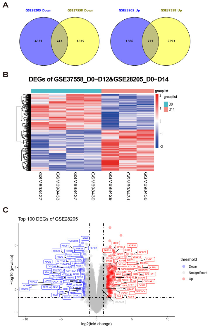
Methods: In this study, we conducted systematic re-analyses of published high-throughput transcriptomic datasets to identify and validate key biological molecules that dynamically regulate MSC osteogenic differentiation. Our approach involved a comprehensive analysis of gene expression patterns across human tissues, followed by the rigorous experimental validation of the identified candidates.

Results: Through integrated analytical and experimental approaches, we utilized high-throughput transcriptomics to identify four critical regulators of MSC osteogenic differentiation: PTBP1, H2AFZ, BCL6, and TTPAL (C20ORF121). Among these, PTBP1 and H2AFZ functioned as positive regulators, while BCL6 and TTPAL acted as negative regulators in osteogenesis. The regulatory roles of these genes in osteogenesis were further validated via overexpression experiments.

Conclusions: Our findings advance our understanding of MSC differentiation fate determination and open new therapeutic possibilities for bone-related disorders. The identification of these regulators provides a foundation for developing targeted interventions in regenerative medicine.

Keywords: MSCs; dynamic regulation; lineage change; osteogenesis; transcriptomic sequencing.


Conflict of interest statement

The authors declare no conflicts of interest.

Figures

Figure 1
Figure 1
Two batches of microarray datasets related to long-term osteogenic induction were analyzed to identify DEGs. (A) Venn diagrams illustrating the intersection of differentially expressed genes (DEGs) between the two microarray datasets. The left panel represents the down-regulated genes, while the right panel represents the up-regulated genes in GSE28205 and GSE37558. The DEGs were identified using thresholds of false discovery rate (FDR) ≤ 0.25 and p-value ≤ 0.05. (B) A heatmap was utilized to visualize the DEGs between the two batches of datasets. (C) The volcano plot in panel C presents the information on the DEGs with a cutoff value of log2(fold change) ≥ 1.5.
Figure 2
Figure 2
Refinement of time-dependent osteogenic induction in MSCs through the analysis of microarray dataset GSE37558. (A) A Venn diagram was generated to illustrate the overlap of DEGs across three time points during osteogenic induction in the GSE37558 dataset. The left panel represents the down-regulated genes, while the right panel represents the up-regulated genes. The DEGs were identified based on thresholds of false discovery rate (FDR) ≤ 0.25 and p-value ≤ 0.05. (B) The DEGs among the three different osteogenic induction time points in the GSE37558 dataset were visualized using a heatmap. (C) The volcano plot in panel C presents the information on the DEGs with a cutoff value of log2(fold change) ≥ 1.5.
Figure 3
Figure 3
Recombining DEGs to enhance the screening and identification of key regulatory molecules. (A,B) Venn diagram showing the intersection of DEGs across time points and microarray datasets from various batches; (C,D) Detailed view of the coexpression network of DEGs, highlighting key genes.
Figure 4
Figure 4
Characterization and multipotent differentiation capacity of mouse bone marrow mesenchymal stem cells (mBMSCs). (A) The flow cytometry analysis of the expression of positive (CD90, CD73, and CD105) and negative (CD34 and CD117) markers of mBMSCs. (B) Alizarin Red S staining for mBMSCs culturing for 21 days in osteogenic medium or basal medium. (C) Oil Red O staining for mBMSCs culturing for 14 days in adipogenic medium or basal medium. (D) Alcian blue staining for mBMSCs culturing for 28 days in chondrogenic medium or basal medium with the method of micromass. Scale bar: 100 μm. All the data are presented as the means ± SEMs. Statistical significance was determined using one-way ANOVA followed by Scheffe’s post hoc test. All the cell experiments were repeated independently in triplicate.
Figure 5
Figure 5
Elucidating the molecular mechanisms governing dynamic osteogenic induction in MSCs through the overexpression of four candidate genes using lentiviral packaging technology. (A) ALP and ARS were performed to evaluate the osteogenic differentiation of BMSCs at 1, 7, 14, and 21 days. Scale bar: 100 μm. (B) A qRT–PCR analysis was conducted to assess the mRNA expression levels of osteogenic markers (ALP, BGLAP, and RUNX2) at 1, 7, 14, and 21 days. (B) A qRT–PCR analysis was conducted to assess the mRNA expression levels of osteogenic markers (ALP, BGLAP, and RUNX2) at 1, 7, 14, and 21 days. (C) The temporal expression of BCL6 and TTPAL mRNA during the osteogenic induction period was analyzed at 1, 7, 14, and 21 days via qRT–PCR. (D) The temporal expression of PTBP1 and H2AFZ mRNA during the osteogenic induction period was analyzed at 1, 7, 14, and 21 days via qRT–PCR. The experiments were performed in triplicate using cells isolated from three separate 2-month-old wild-type C57BL/6J mice. Each replicate was derived from a different mouse. All the data are presented as the means ± SEMs. Statistical significance was determined using one-way ANOVA followed by Scheffe’s post hoc test. * p < 0.05; ** p < 0.01. All the cell experiments were repeated independently in triplicate.
Figure 6
Figure 6
Molecular functions involved in the dynamic regulation of osteogenic induction through the lentiviral-mediated overexpression of four candidate genes. (A) Overexpression efficacy measured by qRT–PCR; (B) the overexpression of BCL6 and TTPAL affects the osteogenic phenotype of BMSCs, as detected by an ARS assay; (C) the overexpression of PTBP1 and H2AFZ alters the osteogenic phenotype of BMSCs, as detected by an ARS assay; (D) flow cytometry analysis showing the effects of PTBP1 and H2AFZ overexpression on apoptosis. Each replicate was derived from a different mouse. All the data are presented as the means ± SEMs. Statistical significance was determined using one-way ANOVA followed by Scheffe’s post hoc test. *** p < 0.001. All the cell experiments were repeated independently in triplicate.

Similar articles

Cited by

References

    1. Yu H., Huang Y., Yang L. Research progress in the use of mesenchymal stem cells and their derived exosomes in the treatment of osteoarthritis. Ageing Res. Rev. 2022;80:101684. doi: 10.1016/j.arr.2022.101684. - DOI - PubMed
    1. Zhang H., Xu R., Li B., Xin Z., Ling Z., Zhu W., Li X., Zhang P., Fu Y., Chen J., et al. LncRNA NEAT1 controls the lineage fates of BMSCs during skeletal aging by impairing mitochondrial function and pluripotency maintenance. Cell Death Differ. 2022;29:351–365. doi: 10.1038/s41418-021-00858-0. - DOI - PMC - PubMed
    1. Han Y., Yang J., Fang J., Zhou Y., Candi E., Wang J., Hua D., Shao C., Shi Y. The secretion profile of mesenchymal stem cells and potential applications in treating human diseases. Signal Transduct. Target. Ther. 2022;7:92. doi: 10.1038/s41392-022-00932-0. - DOI - PMC - PubMed
    1. Wang Y., Fang J., Liu B., Shao C., Shi Y. Reciprocal regulation of mesenchymal stem cells and immune responses. Cell Stem Cell. 2022;29:1515–1530. doi: 10.1016/j.stem.2022.10.001. - DOI - PubMed
    1. Jiang Z., Li N., Shao Q., Zhu D., Feng Y., Wang Y., Yu M., Ren L., Chen Q., Yang G. Light-controlled scaffold- and serum-free hard palatal-derived mesenchymal stem cell aggregates for bone regeneration. Bioeng. Transl. Med. 2023;8:e10334. doi: 10.1002/btm2.10334. - DOI - PMC - PubMed

MeSH terms

Substances

LinkOut - more resources