. 2024 Dec 9;15(12):1583.
doi: 10.3390/genes15121583.

Kir4.1 and Aqp4 Contribution to Schisis Cystic Water Accumulation and Clearance in the Rs1 Exon-1 Del XLRS Rat Model

Affiliations

Kir4.1 and Aqp4 Contribution to Schisis Cystic Water Accumulation and Clearance in the Rs1 Exon-1 Del XLRS Rat Model

Zeljka Smit-McBride et al. Genes (Basel). .

Abstract

Background/objective: The Rs1 exon-1-del rat (Rs1KO) XLRS model shows normal retinal development until postnatal day 12 (P12) when small cystic spaces start to form in the inner nuclear layer. These spaces enlarge rapidly, peak at P15, and then collapse by P19.

Methods: We explored the possible involvement of Kir4.1 and Aqp4, the principal retina channels for water movement and homeostasis, along with Muller glia cells (MGCs), using semi-quantitative fluorescent immunohistochemistry at P7, P9, P12, and P30, in Rs1KO and WT littermates.

Results: Kir4.1 expression was reduced in Rs1KO retinas at all the early time points-P7, P9, and P12-as the schisis cavities began to form; downregulation would reduce water egress from the retina. Aqp4 was upregulated at P30 in Rs1KO retinas during schisis cavity closure but not as cavities formed at P12. When examined by GFAP expression, MGCs were not activated at the preschisis P12 age but showed considerable GFAP expression at P30 following retinal cystic structural damage at P15, indicating that MGCs were activated during the period of retina water removal and cavity closure.

Conclusions: The study results implicate the downregulation of Kir4.1 in schisis formation and a role for both Kir4.1 and Aqp4 upregulation in subsequent schisis closure.

Keywords: Aqp4; Kir4.1; MGC; Muller glia cells; RS1; X-linked retinoschisis; XLRS; aquaporin4; deep capillary plexus; rat retina disease model; retina development; retinoschisin.


Conflict of interest statement

The authors declare no conflicts of interest. The funders had no role in the design of the study; in the collection, analyses, or interpretation of data; in the writing of the manuscript; or in the decision to publish the results.

Figures
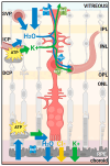
Figure 1
Figure 1
A schematic representation of a retinal Muller glial cell (MGC) illustrating its roles in ions and water drainage from the inner retina toward the retinal vessels. Potassium transport is associated with water drainage through inwardly rectifying potassium channels (Kir4.1) and aquaporin 4 (Aqp4) channels, both located close to the interface of the retinal MGC with retinal vessels and in retinal MGC endfeet at the level of the internal limiting membrane. Image created using BioRender: Smit-McBride, Z. (2024) https://BioRender.com/p88p075 (accessed on 25 November 2024). IPL, inner plexiform layer; INL, inner nuclear layer; OPL, outer plexiform layer; ONL, outer nuclear layer; RPE, retinal pigment epithelium; SVP, superficial vascular plexus; ICP, intermediate capillary plexus; DCP, deep capillary plexus; ATP, adenosine triphosphate.
Figure 2
Figure 2
Retinal schisis occurrence in the rat XLRS animal model. The tissue section of the WT and Rs1KO rat retinas stained with H&E at three time points, showing retinal schisis. H&E stain (hematoxylin and eosin) primarily highlights the nuclei of cells in a blue-purple color while staining the cytoplasm and extracellular matrix a pinkish hue. Time points presented are P7, P15, and P30, in wild type (WT) and Rs1 knockout (Rs1KO). GC, ganglion cells; IPL, inner plexiform layer; INL, inner nuclear layer; OPL, outer plexiform layer; ONL, outer nuclear layer; RPE, retinal pigment epithelium (scale bar = 100 µm).
Figure 3
Figure 3
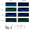
Aquaporin 4 expression and distribution in retinal tissue. Tissue section of WT and Rs1KO rat retinas with Aqp4 fluorescence immunolocalization at various time points: (a) preschisis (P7, P9, and P12) and (b) postschisis (P30). Confocal images show Aqp4 (red) and the nuclear marker DAPI (blue) (c) Expression levels of Aqp4 were analyzed quantitatively using ImageJ software (NIH, Bethesda, MD, USA) in WT and Rs1KO rats: bar graph (left); table (right). The number of replicas was n = 3 for each group (WT and Rs1KO) for P7, P9, and P12, and n = 5 for each group (WT and Rs1KO) for P30. Rs1KO, knockout; SD, standard deviation; WT, wild type. * Statistically significant. *** indicate time gap between P12 (preschisis) and P30 (postschisis). ILM, inner limiting membrane; GCL, ganglion cell layer; IPL, inner plexiform layer; INL, inner nuclear layer; OPL, outer plexiform layer; ONL, outer nuclear layer; RPE, retinal pigment epithelium (scale bar = 100 µ).
Figure 4
Figure 4
Colocalization of Aqp4 expression with perivascular vessels at P30 in WT and Rs1KO rat retinas. The blood vessels (Isolectin B GS-IB4-silver) and Aqp4 (red) are expressed in MC tightly around blood vessels in WT, but in Rs1KO rat retinas, the expression is much more distributed. Confocal images show Aqp4 (in red), the endothelial cell marker Isolectin B, GS-IB4 (silver), and the nuclear marker DAPI (blue). Aqp4 was concentrated in the perivascular (arrows) and inner limiting membrane domains in the WT animals (scale bar = 50 µ).
Figure 5
Figure 5
Kir4.1 expression and distribution in rat retinal tissues. Tissue section of WT and Rs1KO rat retinas with Kir4.1 fluorescence immunolocalization at various time points: (a) preschisis (P7, P9, and P12) and (b) postschisis (P30). Confocal images show Kir4.1 (green) and the nuclear marker DAPI (blue) (c) The expression levels of Kir4.1 were analyzed quantitatively using ImageJ software (NIH, Bethesda, MD, USA) in WT and Rs1KO rats: bar graph (left); table (right). The number of replicas was n = 3 for each group (WT and Rs1KO) for P7, P9, and P12, and n = 5 for each group (WT and Rs1KO) for P30. * Statistically significant. *** indicated time gap between P12 (preschisis) and P30 (postschisis). ILM, inner limiting membrane; GCL, ganglion cell layer; IPL, inner plexiform layer; INL, inner nuclear layer; OPL, outer plexiform layer; ONL, outer nuclear layer; RPE, retinal pigment epithelium (scale bar = 100 µ).
Figure 6
Figure 6
Disruption and thinning of the deep capillary plexus. (a) Retinal flatmounts of WT and Rs1KO rats at P30, with blood vessels labeled for isolectin B (GS-IB4 antibody, silver). (b) Enlarged regions of retinal flatmounts of WT and Rs1KO rats at P30, with blood vessels labeled for isolectin B (GS-IB4 antibody, silver). Aqp 4 (red) showed DCP disruption associated with MGC perivascular processes (scale bar = 60 µ).
Figure 7
Figure 7
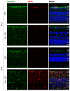
Expression of the MGC marker vimentin and activation marker GFAP. Presented are P12 and P30 time points, both WT and Rs1KO rats. The expression of GFAP and the activation of Muller cells are not present at P12 but are very visible at P30. The number of replicas was n = 3 for each group (WT and Rs1KO) for P12 and P30. Confocal images show GFAP, glial fibrillary activation protein (red), Vim, vimentin (green), and the nuclear marker DAPI (blue). ILM, inner limiting membrane; GCL, ganglion cell layer; IPL, inner plexiform layer; INL, inner nuclear layer; OPL, outer plexiform layer; ONL, outer nuclear layer; OLM, outer limiting membrane (scale bar = 40 µ).
Figure 8
Figure 8
Glutamine synthetase (GS) (silver), exclusively expressed in Muller glia in the retina at P12 and P30, showed the variation in Rs1KO retina MGC soma and nuclei placement, which was spread out and more disorganized in P30 than in P12 images (scale bar = 20 µ).

Similar articles

Cited by

References

    1. Fahim A.T., Ali N., Blachley T., Michaelides M. Peripheral fundus findings in X-linked retinoschisis. Br. J. Ophthalmol. 2017;101:1555–1559. doi: 10.1136/bjophthalmol-2016-310110. - DOI - PubMed
    1. Rao P., Dedania V.S., Drenser K.A. Congenital X-Linked Retinoschisis: An Updated Clinical Review. Asia Pac. J. Ophthalmol. 2018;7:169–175. doi: 10.22608/201803. - DOI - PubMed
    1. Chen D., Xu T., Tu M., Xu J., Zhou C., Cheng L., Yang R., Yang T., Zheng W., He X., et al. Recapitulating X-Linked Juvenile Retinoschisis in Mouse Model by Knock-In Patient-Specific Novel Mutation. Front. Mol. Neurosci. 2017;10:453. doi: 10.3389/fnmol.2017.00453. - DOI - PMC - PubMed
    1. Kwon H.J., Kim Y.N., Min C.H., Kim Y.J., Lee J., Lee J.Y., Yoon Y.H. MACULAR MICROVASCULATURE IN X-LINKED RETINOSCHISIS: Optical Coherence Tomography and Optical Coherence Tomography Angiography Study. Retina. 2022;42:1939–1949. doi: 10.1097/IAE.0000000000003539. - DOI - PubMed
    1. Romano F., Arrigo A., Ch’ng S.W., Battaglia Parodi M., Manitto M.P., Martina E., Bandello F., Stanga P.E. Capillary Network Alterations in X-Linked Retinoschisis Imaged on Optical Coherence Tomography Angiography. Retina. 2019;39:1761–1767. doi: 10.1097/IAE.0000000000002222. - DOI - PubMed