Review
. 2024 Dec 13;15(12):1596.
doi: 10.3390/genes15121596.

Exploring the Molecular Link Between Diabetes and Erectile Dysfunction Through Single-Cell Transcriptome Analysis

Affiliations
Review

Exploring the Molecular Link Between Diabetes and Erectile Dysfunction Through Single-Cell Transcriptome Analysis

Mahmuda Begum et al. Genes (Basel). .

Abstract

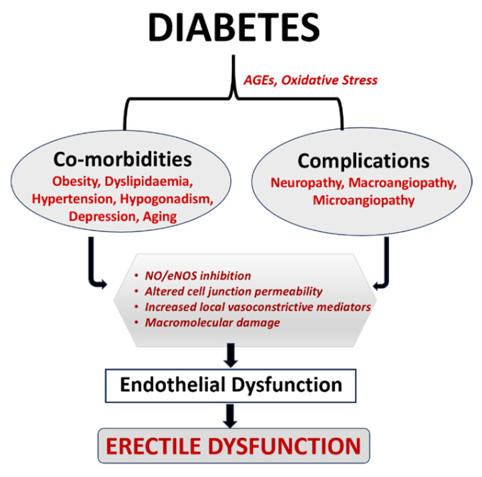
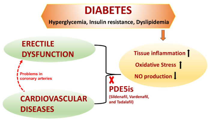
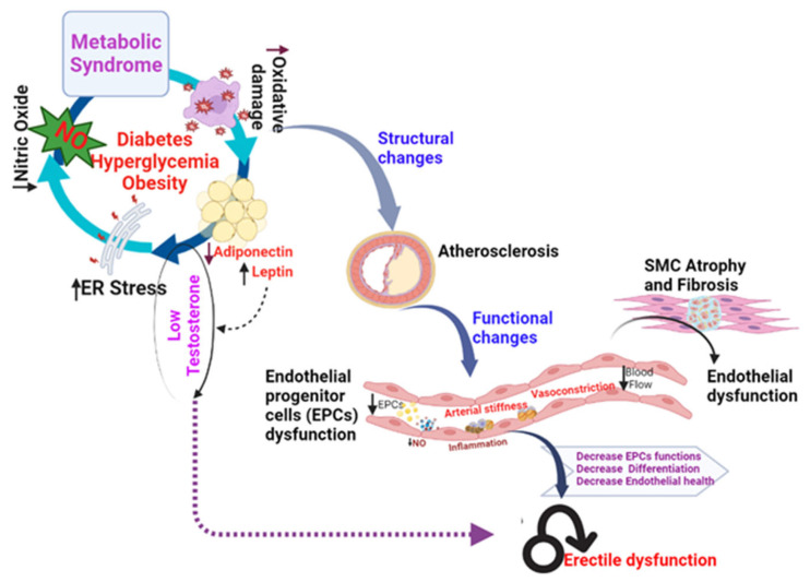
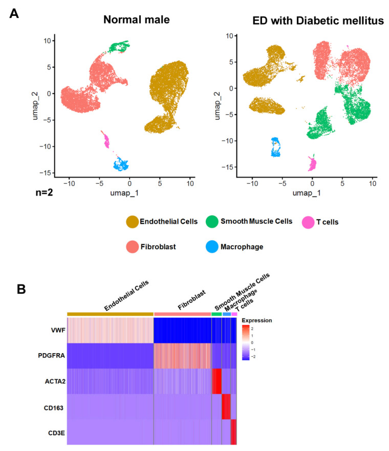
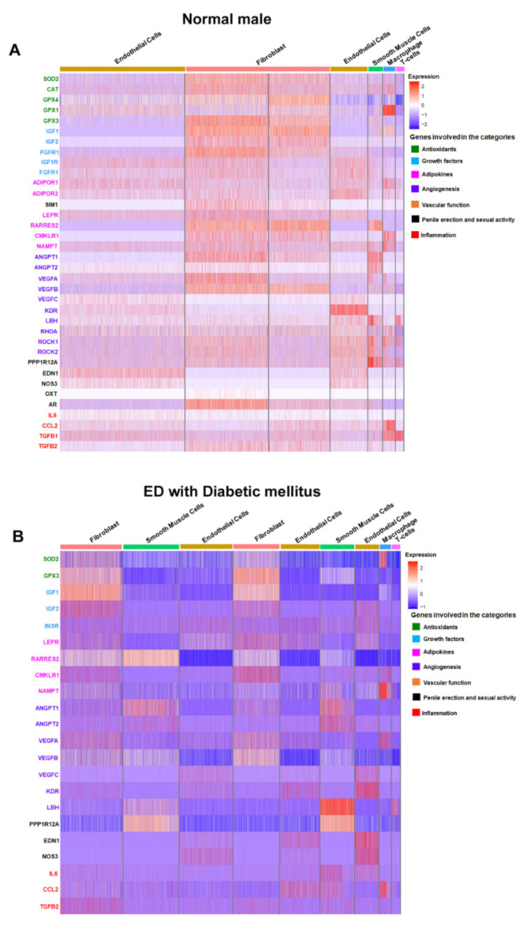
Erectile dysfunction (ED) is a pathophysiological condition in which the patients cannot achieve an erection during sexual activity, and it is often overlooked yet prevalent among diabetic men, globally affecting approximately 35-75% of diabetic individuals. The precise mechanisms through which diabetes contributes to ED remain elusive, but the existing literature suggests the potential involvement of nerve and vascular damage that affects the penile supply. In the present review, we reanalyze the existing human single-cell transcriptomic data from patients having diabetes mellitus-associated ED with normal erections. The analysis validates the expression of genes associated with antioxidative pathways, growth factors, adipokines, angiogenesis, vascular functions, penile erection, sexual function, and inflammation in diverse cell types from healthy individuals and those with ED. Our transcriptomic analysis reveals alterations in the expression of adiponectin receptors in the pathogenesis of ED compared to their counterparts in healthy subjects. This comprehensive review sheds light on the molecular underpinnings of ED in the context of diabetes, providing an in-depth understanding of the biological and cellular alterations involved and paving the way for possible targeted therapeutic discoveries in the field of diabetes-associated male infertility.

Keywords: diabetes; diabetic sexual health; erectile dysfunction; human single-cell transcriptome data; men’s sexual health; microvascular complications; single-cell analysis.


Conflict of interest statement

The authors declare no conflict of interest.

Figures

Figure 1
Figure 1
Association of Diabetes and Emergency Department: The hyperglycemic stage of diabetes causes oxidative stress and AGE formation, which further leads to a variety of comorbidities and complications. The aforementioned problems also cause endothelial dysfunction-induced ED through increased local vasoconstrictive mediators, altered cell junction permeability, inhibition of NO/eNOS, and macromolecular damage [18,19].
Figure 2
Figure 2
Treatment of diabetes-associated ED. Hyperglycemia, insulin resistance, and dyslipidemia related to diabetes cause oxidative stress, tissue inflammation, and decreased NO production. Commercially available PDE5is, such as Vardenafil, Tadalafil, and Sildenafil, can mitigate this stress-related mechanism, making them an excellent treatment approach for CVD and ED. PDE5is, phosphodiesterase 5 inhibitors; CVD, cardiovascular diseases; ED, erectile dysfunction [31].
Figure 3
Figure 3
Factors contributing to erectile dysfunction (ED). Endothelial dysfunction leading to ED is attributed to factors such as obesity, stress of the endoplasmic reticulum (ER), and oxidative stress.
Figure 4
Figure 4
Cellular Composition Analysis in Normal and Erectile Dysfunction (ED) Tissues with Diabetes Mellitus. (A) Uniform Manifold Approximation and Projection (UMAP) visualization of single-cell RNA sequencing data from human corpus cavernosum penile tissues comparing a normal male (left) and an ED patient with diabetes mellitus (right). Each dot represents a single cell, color-coded by cell type: endothelial cells (yellow), smooth muscle cells (green), T cells (blue), fibroblasts (pink), and macrophages (red). The number of cells analyzed (n) is indicated below each plot. (B) Heatmap depicting the expression profile of selected marker genes across the identified cell types from both conditions. Rows represent individual genes and columns represent cell types as indicated at the top. Color intensity reflects the level of gene expression, with blue indicating low expression and red indicating high expression. Marker genes are VWF for endothelial cells, PDGFRA for fibroblasts, ACTA2 for smooth muscle cells, CD163 for macrophages, and CD3E for T cells. Data were analyzed using the software Seurat v4.1.1 implemented in R v4.2.1.
Figure 5
Figure 5
Differential Gene Expression in Penile Tissue of Normal and ED-Affected Males with Diabetes Mellitus. (A) Heatmap of gene expression profiles in human corpus cavernosum penile tissue from a normal male. The rows represent individual genes involved in various biological processes such as antioxidants, growth factors, adipokines, angiogenesis, vascular function, penile erection and sexual activity, and inflammation, as color-coded on the right. Columns represent cell types: endothelial cells and fibroblasts. Expression levels are indicated by the color gradient, with red representing high expression, white representing neutral, and blue indicating low expression. (B) Comparative heatmap of gene expression profiles in human corpus cavernosum penile tissue from an ED patient with diabetes mellitus. Layout and color-coding are similar to (A), showing the same genes and biological process categories across different cell types: endothelial cells, smooth muscle cells, fibroblasts, and macrophages/immune cells.

Similar articles

References

    1. CDC Global Health—Infographics—World Diabetes Day. [(accessed on 3 October 2023)];2020 Available online: https://www.cdc.gov/globalhealth/infographics/diabetes/world-diabetes-da....
    1. Diabetes PAHO/WHO: Pan American Health Organization. [(accessed on 8 December 2023)]. Available online: https://www.paho.org/en/topics/diabetes.
    1. Mohiuddin M.S., Himeno T., Inoue R., Miura-Yura E., Yamada Y., Nakai-Shimoda H., Asano S., Kato M., Motegi M., Kondo M., et al. Glucagon-Like Peptide-1 Receptor Agonist Protects Dorsal Root Ganglion Neurons against Oxidative Insult. J. Diabetes Res. 2019;2019:9426014. doi: 10.1155/2019/9426014. - DOI - PMC - PubMed
    1. Mohiuddin M.S., Himeno T., Yamada Y., Morishita Y., Kondo M., Tsunekawa S., Kato Y., Nakamura J., Kamiya H. Glucagon Prevents Cytotoxicity Induced by Methylglyoxal in a Rat Neuronal Cell Line Model. Biomolecules. 2021;11:287. doi: 10.3390/biom11020287. - DOI - PMC - PubMed
    1. Cosentino F., Assenza G.E. Diabetes and Inflammation. Herz. 2004;29:749–759. doi: 10.1007/s00059-004-2635-8. - DOI - PubMed

LinkOut - more resources