. 2024 Dec 19;15(12):1623.
doi: 10.3390/genes15121623.

Side- and Disease-Dependent Changes in Human Aortic Valve Cell Population and Transcriptomic Heterogeneity Determined by Single-Cell RNA Sequencing

Affiliations

Side- and Disease-Dependent Changes in Human Aortic Valve Cell Population and Transcriptomic Heterogeneity Determined by Single-Cell RNA Sequencing

Nicolas Villa-Roel et al. Genes (Basel). .

Abstract

Background: Calcific aortic valve disease (CAVD) is a highly prevalent disease, especially in the elderly population, but there are no effective drug therapies other than aortic valve repair or replacement. CAVD develops preferentially on the fibrosa side, while the ventricularis side remains relatively spared through unknown mechanisms. We hypothesized that the fibrosa is prone to the disease due to side-dependent differences in transcriptomic patterns and cell phenotypes.

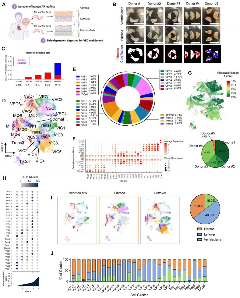
Methods: To test this hypothesis, we performed single-cell RNA sequencing using a new method to collect endothelial-enriched samples independently from the fibrosa and ventricularis sides of freshly obtained human aortic valve leaflets from five donors, ranging from non-diseased to fibrocalcific stages.

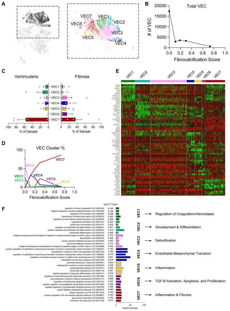
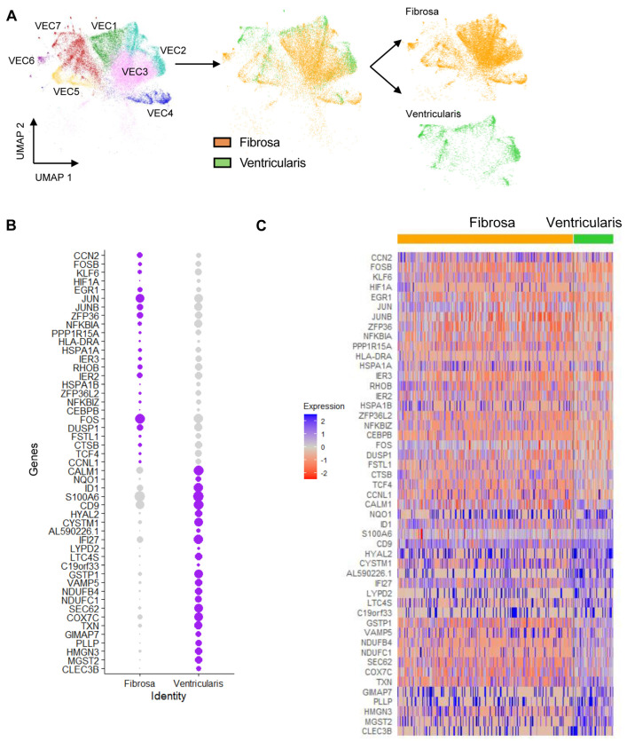
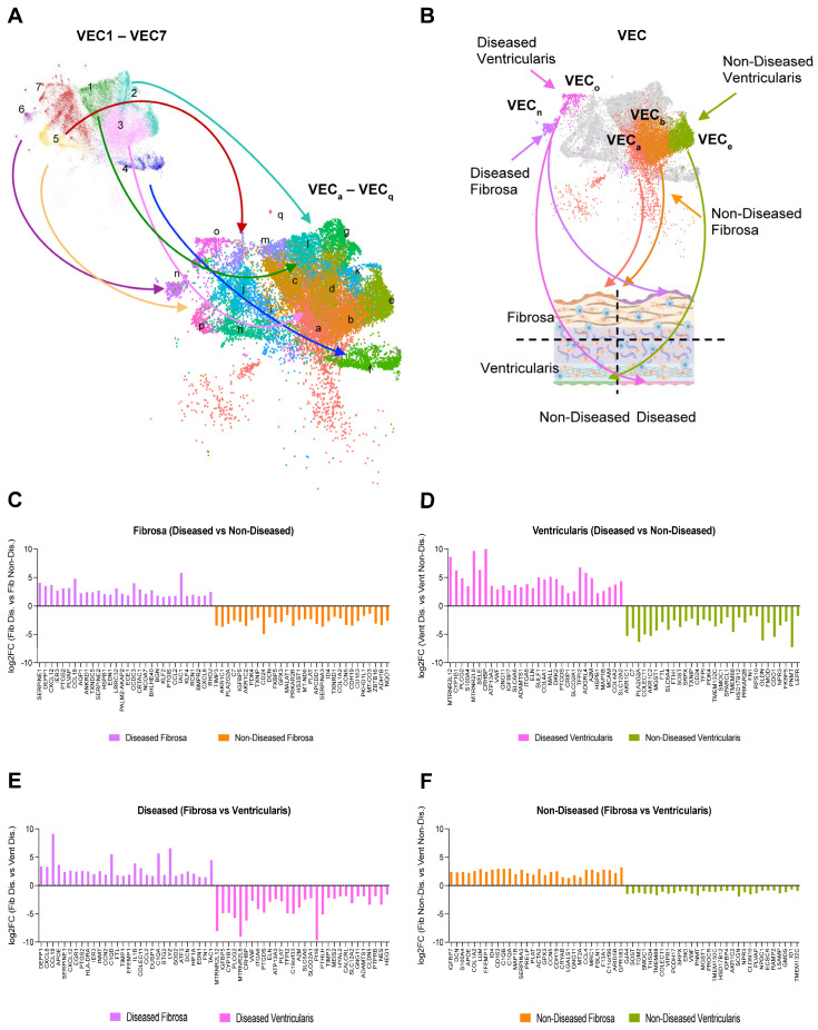
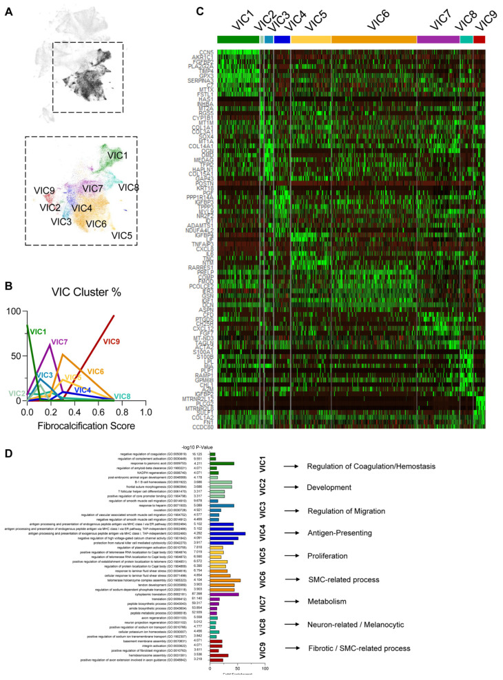
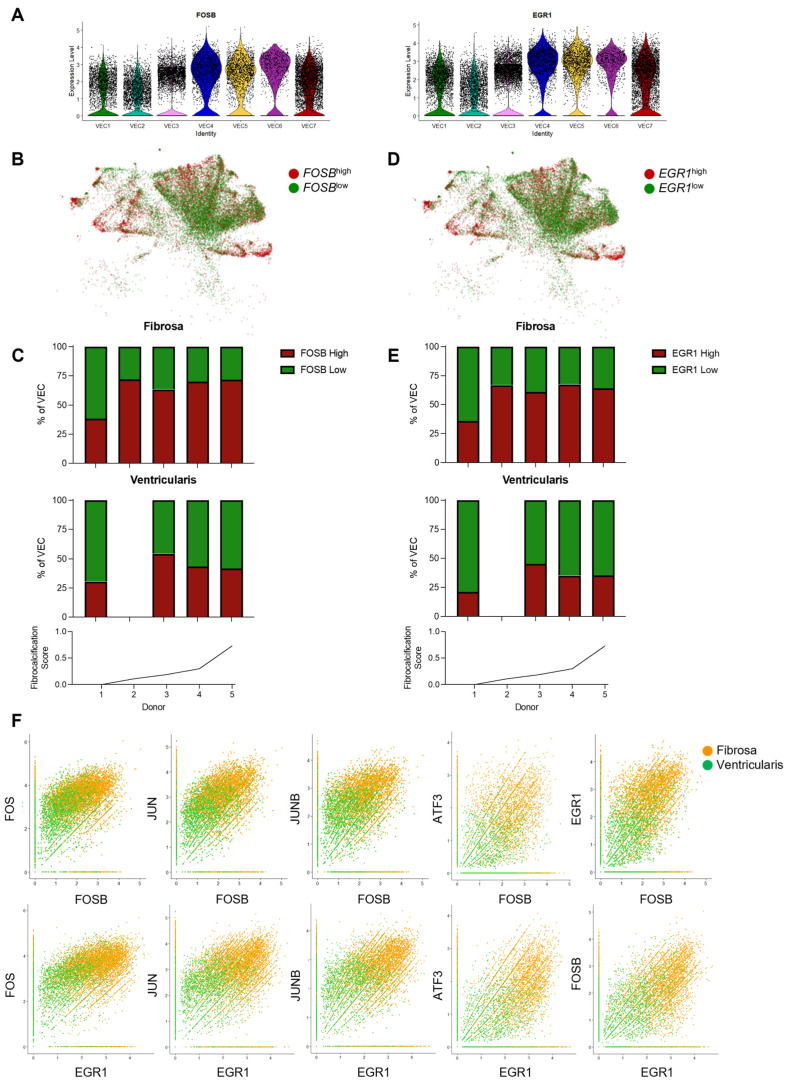
Results: From the 82,356 aortic valve cells analyzed, we found 27 cell clusters, including seven valvular endothelial cell (VEC), nine valvular interstitial cell (VIC), and seven immune, three transitional, and one stromal cell population. We identified several side-dependent VEC subtypes with unique gene expression patterns. Homeostatic VIC clusters were abundant in non-diseased tissues, while VICs enriched with fibrocalcific genes and pathways were more prevalent in diseased leaflets. Furthermore, homeostatic macrophage (MΦ) clusters decreased while inflammatory MΦ and T-cell clusters increased with disease progression. A foamy MΦ cluster was increased in the fibrosa of mildly diseased tissues. Some side-dependent VEC clusters represented non-diseased, protective phenotypes, while others were CAVD-associated and were characterized by genes enriched in pathways of inflammation, endothelial-mesenchymal transition, apoptosis, proliferation, and fibrosis. Interestingly, we found several activator protein-1 (AP-1)-related transcription factors (FOSB, FOS, JUN, JUNB) and EGR1 to be upregulated in the fibrosa and diseased aortic valve leaflets.

Conclusions: Our results showed that VECs are highly heterogeneous in a side- and CAVD-dependent manner. Unique VEC clusters and their differentially regulated genes and pathways found in the fibrosa of diseased tissues may represent novel pathogenic mechanisms and potential therapeutic targets.

Keywords: AP-1 related transcription factors; aortic sclerosis; aortid stenosis; calcific aortic valve disease; endothelial-to-mesenchymal transition; human aortic valves; inflammation; single-cell RNA sequencing; transcriptomics; valve endothelial cells; valve interstitial cells.


Conflict of interest statement

H.J. is the founder of Flokines Pharma. All other authors report no conflicts of interest.

Figures

Figure 1
Figure 1
Side-dependent, VEC-enriched scRNAseq of human aortic valve reveals 27 cell clusters that vary with fibrocalcific stage: (A) Strategy for side-dependent, VEC-enriched scRNAseq of human aortic valve leaflets. (B) Gross images of aortic valve leaflets used for scRNAseq taken from ventricularis (top) and fibrosa (middle) views. The bottom row shows quantified segmentation images of the fibrotic (red) and calcified (blue) regions. Scale bar = 1 cm. (C) Quantification of fibrocalcification score for each donor. (D) UMAP of the 82,356 cells from the merged and filtered dataset, with labeled cell clusters. (E) Proportions of cell types (VECs, VICs, transitional, and immune) in the dataset. (F) Dot plot depicting average expression of annotation markers for each cell cluster. (G) Labeling of each cell by fibrocalcification score and proportion of cells per donor in the dataset. (H) Proportion of each cell cluster based on donor fibrocalcification score. (I) UMAP analysis of cell clusters from ventricularis, fibrosa, and leftover (mixed), and proportion of cells from each sample. (J) Side-dependent proportions in each cell cluster. VEC: valvular endothelial cell; scRNAseq: single-cell RNA sequencing; VIC: valvular interstitial cell; UMAP: uniform manifold approximation and projection.
Figure 2
Figure 2
VEC clustering patterns change with disease stage: (A) UMAP of the 7 VEC clusters. (B) Total VEC numbers decrease with disease progression. (C) VEC cluster proportions in ventricularis vs. fibrosa. (D) VEC cluster proportions change as a function of disease stage. (E) Heatmap highlighting the top 10 most enriched genes in each VEC cluster. (F) Top 5 significantly enriched biological processes from a Gene Ontology analysis using the top 200 upregulated genes in each VEC cluster and a representative process for each cluster.
Figure 3
Figure 3
Identification of side-dependent VEC clustering patterns and differentially expressed genes (DEGs): (A) Side-dependent separation of VEC UMAP into fibrosa vs. ventricularis. (B) Side-dependent expression of DEGs in fibrosa or ventricularis VECs. (C) Heatmap classifying side-dependent DEGs in fibrosa or ventricularis VECs. Genes upregulated in the fibrosa of diseased tissues and genes upregulated in the ventricularis of non-calcified tissues are indicated.
Figure 4
Figure 4
Identification of side- and disease-dependent sub-clusters of VECs and genes: (A) Seven VEC clusters (VEC1–7) were divided into 17 sub-clusters (VECa–q). (B) Identification of diseased or non-diseased, side-dependent sub-clusters of VECs (non-diseased fibrosa = sub-clusters a and b, non-diseased ventricularis = sub-cluster e, diseased fibrosa = sub-cluster n, and diseased ventricularis = sub-cluster o). (C,D) Top 30 DEGs between diseased and non-diseased VEC in the fibrosa (C) and ventricularis (D) sub-clusters. (E,F) Top 30 DEGs between fibrosa and ventricularis VECs in diseased (E) and non-diseased (F) sub-clusters.
Figure 5
Figure 5
VIC clustering patterns change with disease stage: (A) UMAP of the 9 VIC clusters. (B) VIC cluster proportions change as a function of disease stage. (C) Heatmap highlighting the top 10 most enriched genes in each VIC cluster. (D) Top 5 significantly enriched biological processes from a Gene Ontology analysis using the top 200 upregulated genes in each VIC cluster and a representative process for each cluster.
Figure 6
Figure 6
FOSB and EGR1 are upregulated in fibrosa VECs in diseased aortic valve leaflets: (A) Violin plots showing that FOSB and EGR1 expression is increased in disease-associated VEC clusters. (B) VEC UMAP separated by high FOSB expression or low FOSB expression (average relative FOSB expression = 1.5). (C) Disease-stage-associated proportions of FOSBhigh and FOSBlow VECs, split in a side-dependent manner. (D) UMAP of the VECs separated by high EGR1 expression or low EGR1 expression (average relative EGR1 expression = 1.5). (E) Stacked bar graph of individual donor proportions of EGR1high and EGR1low VECs, split by layer of origin. (F) VECs from the fibrosa (orange) and ventricularis (green) show co-expression of FOSB (top row) or EGR1 (bottom row) with other AP-1-related transcription factors (FOS, JUN, JUNB, ATF3, FOSB, and EGR1).

Similar articles

References

    1. Virani S.S., Alonso A., Aparicio H.J., Benjamin E.J., Bittencourt M.S., Callaway C.W., Carson A.P., Chamberlain A.M., Cheng S., Delling F.N., et al. Heart disease and stroke statistics-2021 update: A report from the american heart association. Circulation. 2021;143:e254–e743. - PubMed
    1. Yadgir S., Johnson C.O., Aboyans V., Adebayo O.M., Adedoyin R.A., Afarideh M., Alahdab F., Alashi A., Alipour V., Arabloo J., et al. Global, regional, and national burden of calcific aortic valve and degenerative mitral valve diseases, 1990–2017. Circulation. 2020;141:1670–1680. doi: 10.1161/CIRCULATIONAHA.119.043391. - DOI - PubMed
    1. Kraler S., Blaser M.C., Aikawa E., Camici G.G., Luscher T.F. Calcific aortic valve disease: From molecular and cellular mechanisms to medical therapy. Eur. Heart J. 2022;43:683–697. doi: 10.1093/eurheartj/ehab757. - DOI - PMC - PubMed
    1. Otto C.M., Nishimura R.A., Bonow R.O., Carabello B.A., Erwin J.P., 3rd, Gentile F., Jneid H., Krieger E.V., Mack M., McLeod C., et al. 2020 ACC/AHA guideline for the management of patients with valvular heart disease: A report of the American College of Cardiology/American Heart Association Joint Committee on Clinical Practice Guidelines. Circulation. 2021;143:e72–e227. - PubMed
    1. Nsaibia M.J., Devendran A., Goubaa E., Bouitbir J., Capoulade R., Bouchareb R. Implication of lipids in calcified aortic valve pathogenesis: Why did statins fail? J. Clin. Med. 2022;11:3331. doi: 10.3390/jcm11123331. - DOI - PMC - PubMed

Supplementary concepts