Review
. 2024 Dec 20;15(12):1632.
doi: 10.3390/genes15121632.

Zonulopathies as Genetic Disorders of the Extracellular Matrix

Affiliations
Review

Zonulopathies as Genetic Disorders of the Extracellular Matrix

Chimwemwe Chipeta et al. Genes (Basel). .

Abstract

The zonular fibres are formed primarily of fibrillin-1, a large extracellular matrix (ECM) glycoprotein, and also contain other constituents such as LTBP-2, ADAMTSL6, MFAP-2 and EMILIN-1, amongst others. They are critical for sight, holding the crystalline lens in place and being necessary for accommodation. Zonulopathies refer to conditions in which there is a lack or disruption of zonular support to the lens and may clinically be manifested as ectopia lens (EL)-defined as subluxation of the lens outside of the pupillary plane or frank displacement (dislocation) into the vitreous or anterior segment. Genes implicated in EL include those intimately involved in the formation and function of these glycoproteins as well as other genes involved in the extracellular matrix (ECM). As such, genetic pathogenic variants causing EL are primarily disorders of the ECM, causing zonular weakness by (1) directly affecting the protein components of the zonule, (2) affecting proteins involved in the regulation of zonular formation and (3) causing the dysregulation of ECM components leading to progressive zonular weakness. Herein, we discuss the clinical manifestations of zonulopathy and the underlying pathogenetic mechanisms.

Keywords: Marfan syndrome; ciliary zonule; ectopia lentis; zonulopathy.


Conflict of interest statement

The authors declare no conflict of interest.

Figures
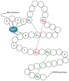
Figure 1
Figure 1
Illustration of a cbEGF-like domain in the fibrillin-1 protein. This is a possible representation of how a fibrillin-1 cbEGF-like domain could look like based on an amino acid sequence and previous published studies [16,17,18]. Calcium (Ca2+) is found bound to several amino acids in the calcium-binding region in the cbEGF-like #35 domain, and shows the conserved amino acids for this domain. Disulphide bonds are represented with coloured dotted lines and show the 3 cysteine pairs [C1–C3 (blue), C2–C4 (red) and C5–C6 (green)].
Figure 2
Figure 2
Structure of ADAMTS superfamily proteins implicated in ectopia lentis. ADAMTS proteins share a structurally similar N-terminal proteinase domain and differ in their C-terminal ancillary domain. ADAMTS-like (ADAMTSL) proteins are structurally similar to ADAMTS proteins but lack the catalytic domains.
Figure 3
Figure 3
Summary of the interactions between ADAMTS proteins and fibrillin-1.
Figure 4
Figure 4
Summary of important proteins implicated in the ectopia lentis phenotype. Picture representing part of the lens on the left and part of the ciliary body on the right. The lines between the lens and ciliary body represent the zonular fibres. Note that the zonular fibres are composed primarily of fibrillin-1. The protein names are placed either in a circle (with number) or a square (names): LTBP-2 = latent TGFβ-binding protein 2; XVIII = collagen XVIII; 10 = ADAMTS10; 17 = ADAMTS17; 18 = ADAMTS18; L4 = ADAMTSL4; XFM = pseudoexfoliative material.

Similar articles

References

    1. De Maria A., Wilmarth P.A., David L.L., Bassnett S. Proteomic Analysis of the Bovine and Human Ciliary Zonule. Investig. Opthalmol. Vis. Sci. 2017;58:573–585. doi: 10.1167/iovs.16-20866. - DOI - PMC - PubMed
    1. Loeys B.L., Dietz H.C., Braverman A.C., Callewaert B.L., De Backer J., Devereux R.B., Hilhorst-Hofstee Y., Jondeau G., Faivre L., Milewicz D.M., et al. The revised Ghent nosology for the Marfan syndrome. J. Med. Genet. 2010;47:476–485. doi: 10.1136/jmg.2009.072785. - DOI - PubMed
    1. Guo D., Li S., Xiao X., Jiang Y., Wang Y., Jin G., Wang J., Ouyang J., Jia X., Sun W., et al. Clinical and Genetic Landscape of Ectopia Lentis Based on a Cohort of Patients From 156 Families. Investig. Opthalmol. Vis. Sci. 2024;65:20. doi: 10.1167/iovs.65.1.20. - DOI - PMC - PubMed
    1. Mönestam E.I. Incidence of Dislocation of Intraocular Lenses and Pseudophakodonesis 10 Years after Cataract Surgery. Ophthalmology. 2009;116:2315–2320. doi: 10.1016/j.ophtha.2009.05.015. - DOI - PubMed
    1. Dabrowska-Kloda K., Kloda T., Boudiaf S., Jakobsson G., Stenevi U. Incidence and risk factors of late in-the-bag intraocular lens dislocation: Evaluation of 140 eyes between 1992 and 2012. J. Cataract. Refract. Surg. 2015;41:1376–1382. doi: 10.1016/j.jcrs.2014.10.040. - DOI - PubMed

MeSH terms

LinkOut - more resources