. 2024 Dec 20;13(24):4134.
doi: 10.3390/foods13244134.

Assessment of Immobilized Lacticaseibacillus rhamnosus OLXAL-1 Cells on Oat Flakes for Functional Regulation of the Intestinal Microbiome in a Type-1 Diabetic Animal Model

Affiliations

Assessment of Immobilized Lacticaseibacillus rhamnosus OLXAL-1 Cells on Oat Flakes for Functional Regulation of the Intestinal Microbiome in a Type-1 Diabetic Animal Model

Grigorios Nelios et al. Foods. .

Abstract

The aim of this study was to examine the effect of free or immobilized Lacticaseibacillus rhamnosus OLXAL-1 cells on oat flakes on the gut microbiota and metabolic and inflammatory markers in a streptozotocin (STZ)-induced Type-1 Diabetes Mellitus (T1DM) animal model. Forty-eight male Wistar rats were assigned into eight groups (n = 6): healthy or diabetic animals that received either a control diet (CD and DCD), an oat-supplemented diet (OD and DOD), a diet supplemented with free L. rhamnosus OLXAL-1 cells (CFC and DFC), or a diet supplemented with immobilized L. rhamnosus OLXAL-1 cells on oat flakes (CIC and DIC). Neither L. rhamnosus OLXAL-1 nor oat supplementation led to any significant positive effects on body weight, insulin levels, plasma glucose concentrations, or lipid profile parameters. L. rhamnosus OLXAL-1 administration resulted in a rise in the relative abundances of Lactobacillus and Bifidobacterium, as well as increased levels of lactic, acetic, and butyric acids in the feces of the diabetic animals. Additionally, supplementation with oat flakes significantly reduced the microbial populations of E. coli, Enterobacteriaceae, coliforms, staphylococci, and enterococci and lowered IL-1β levels in the blood plasma of diabetic animals. These findings suggested that probiotic food-based strategies could have a potential therapeutic role in managing dysbiosis and inflammation associated with T1DM.

Keywords: cell immobilization; gut microbiome; oat flakes; presumptive probiotic; type-1 diabetes mellitus.


Conflict of interest statement

The authors declare no conflicts of interest.

Figures

Figure 1
Figure 1
Body weight of control and diabetic rats over the 4-week dietary intervention period. The values are presented as mean ± SD (n = 6 per group). Significant differences (p < 0.001) among animal groups are indicated by different letters. CD: control animals following the control diet; OD: control animals following the oat flakes diet; CIC: control animals following the diet enriched with immobilized L. rhamnosus OLXAL-1 cells on oat flakes; CFC: control animals following the diet enriched with free L. rhamnosus OLXAL-1 cells; DCD: diabetic animals following the control diet; DOD: diabetic animals following the oat flakes diet; DIC: diabetic animals following the diet enriched with immobilized L. rhamnosus OLXAL-1 cells on oat flakes; DFC: diabetic animals following the diet enriched with free L. rhamnosus OLXAL-1 cells.
Figure 2
Figure 2
Plasma glucose concentrations of control and diabetic rats at the beginning (week 0) and the end (week 4) of the 4-week dietary protocol. The values are presented as mean ± SD (n = 6 per group). Significant differences (p < 0.001) among animal groups are indicated by different letters. CD: control animals following the control diet; OD: control animals following the oat flakes diet; CIC: control animals following the diet enriched with immobilized L. rhamnosus OLXAL-1 cells on oat flakes; CFC: control animals following the diet enriched with free L. rhamnosus OLXAL-1 cells; DCD: diabetic animals following the control diet; DOD: diabetic animals following the oat flakes diet; DIC: diabetic animals following the diet enriched with immobilized L. rhamnosus OLXAL-1 cells on oat flakes; DFC: diabetic animals following the diet enriched with free L. rhamnosus OLXAL-1 cells.
Figure 3
Figure 3
Plasma concentrations of (a) insulin (ng/mL) at the start and end of the dietary intervention, and (b) TNF-α (pg/mL), (c) IL-6 (pg/mL), and (d) IL-1β (pg/mL) at the end of the dietary intervention in control and diabetic rats. The values are presented as mean ± SD (n = 6 per group). Significant differences (p < 0.001) among animal groups are indicated by different letters. CD: control animals following the control diet; OD: control animals following the oat flakes diet; CIC: control animals following the diet enriched with immobilized L. rhamnosus OLXAL-1 cells on oat flakes; CFC: control animals following the diet enriched with free L. rhamnosus OLXAL-1 cells; DCD: diabetic animals following the control diet; DOD: diabetic animals following the oat flakes diet; DIC: diabetic animals following the diet enriched with immobilized L. rhamnosus OLXAL-1 cells on oat flakes; DFC: diabetic animals following the diet enriched with free L. rhamnosus OLXAL-1 cells.
Figure 4
Figure 4
Plasma concentrations of (a) total cholesterol (mg/dL), (b) triglycerides (mg/dL), (c) HDL-cholesterol (mg/dL), (d) LDL-cholesterol (mg/dL), (e) SGPT/ALT (U/L), and (f) SGOT/AST (U/L) in control and diabetic rats at the beginning (week 0) and the end (week 4) of the 4-week dietary protocol. The values are presented as mean ± SD (n = 6 per group). Significant differences (p < 0.05) among animal groups are indicated by different letters. CD: control animals following the control diet; OD: control animals following the oat flakes diet; CIC: control animals following the diet enriched with immobilized L. rhamnosus OLXAL-1 cells on oat flakes; CFC: control animals following the diet enriched with free L. rhamnosus OLXAL-1 cells; DCD: diabetic animals following the control diet; DOD: diabetic animals following the oat flakes diet; DIC: diabetic animals following the diet enriched with immobilized L. rhamnosus OLXAL-1 cells on oat flakes; DFC: diabetic animals following the diet enriched with free L. rhamnosus OLXAL-1 cells.
Figure 5
Figure 5
Effect of the dietary intervention on fecal microbiota populations in both control and diabetic rats at (a) the beginning and (b) the end of the 4-week protocol. The values are presented as mean ± SD (n = 6 per group). Uppercase letters A, B, and C, respectively, denote overall health condition effect, overall diet effect, and interaction between health condition and diet type. * p < 0.05; ** p < 0.01; *** p < 0.001. Significant differences (p < 0.05) among animal groups for each bacteria population within the same time point are indicated by lowercase different letters. CD: control animals following the control diet; OD: control animals following the oat flakes diet; CIC: control animals following the diet enriched with immobilized L. rhamnosus OLXAL-1 cells on oat flakes; CFC: control animals following the diet enriched with free L. rhamnosus OLXAL-1 cells; DCD: diabetic animals following the control diet; DOD: diabetic animals following the oat flakes diet; DIC: diabetic animals following the diet enriched with immobilized L. rhamnosus OLXAL-1 cells on oat flakes; DFC: diabetic animals following the diet enriched with free L. rhamnosus OLXAL-1 cells.
Figure 6
Figure 6
Effect of the dietary intervention on Shannon and Simpson index values in fecal samples of healthy and diabetic rats, as determined by 16S rRNA NGS at the beginning (w0) and the end (w4) of the 4-week protocol. CD: control animals following the control diet; OD: control animals following the oat flakes diet; CIC: control animals following the diet enriched with immobilized L. rhamnosus OLXAL-1 cells on oat flakes; CFC: control animals following the diet enriched with free L. rhamnosus OLXAL-1 cells; DCD: diabetic animals following the control diet; DOD: diabetic animals following the oat flakes diet; DIC: diabetic animals following the diet enriched with immobilized L. rhamnosus OLXAL-1 cells on oat flakes; DFC: diabetic animals following the diet enriched with free L. rhamnosus OLXAL-1 cells.
Figure 7
Figure 7
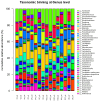
Effect of the dietary intervention on the genus-level taxonomic binning with normalized relative abundances (%) in the feces of control and diabetic rats, as determined through 16S rRNA NGS at the beginning (w0) and the end (w4) of the 4-week protocol (n = 2 per group). CD: control animals following the control diet; OD: control animals following the oat flakes diet; CIC: control animals following the diet enriched with immobilized L. rhamnosus OLXAL-1 cells on oat flakes; CFC: control animals following the diet enriched with free L. rhamnosus OLXAL-1 cells; DCD: diabetic animals following the control diet; DOD: diabetic animals following the oat flakes diet; DIC: diabetic animals following the diet enriched with immobilized L. rhamnosus OLXAL-1 cells on oat flakes; DFC: diabetic animals following the diet enriched with free L. rhamnosus OLXAL-1 cells.

References

    1. Furuyama K., Chera S., van Gurp L., Oropeza D., Ghila L., Damond N., Vethe H., Paulo J.A., Joosten A.M., Berney T., et al. Diabetes relief in mice by glucose-sensing insulin-secreting human α-cells. Nature. 2019;567:43–48. doi: 10.1038/s41586-019-0942-8. - DOI - PMC - PubMed
    1. Rawshani A., Landin-Olsson M., Svensson A.M., Nyström L., Arnqvist H.J., Bolinder J., Gudbjörnsdottir S. The incidence of diabetes among 0-34 year olds in Sweden: New data and better methods. Diabetologia. 2014;57:1375–1381. doi: 10.1007/s00125-014-3225-9. - DOI - PMC - PubMed
    1. Diaz-Valencia P.A., Bougnères P., Valleron A.J. Global epidemiology of type 1 diabetes in young adults and adults: A systematic review. BMC Public Health. 2015;15:255. doi: 10.1186/s12889-015-1591-y. - DOI - PMC - PubMed
    1. Atkinson M.A. The pathogenesis and natural history of type 1 diabetes. Cold Spring Harb. Perspect. Med. 2012;2:a007641. doi: 10.1101/cshperspect.a007641. - DOI - PMC - PubMed
    1. Niechciał E., Michalak M., Skowrońska B., Fichna P. Increasing trend of childhood type 1 diabetes incidence: 20-year observation from Greater Poland Province, Poland. Acta Diabetol. 2024;61:1609–1617. doi: 10.1007/s00592-024-02339-5. - DOI - PMC - PubMed

LinkOut - more resources