. 2024 Nov 22;12(12):2669.
doi: 10.3390/biomedicines12122669.

Age-Related Choroidal Involution Is Associated with the Senescence of Endothelial Progenitor Cells in the Choroid

Affiliations

Age-Related Choroidal Involution Is Associated with the Senescence of Endothelial Progenitor Cells in the Choroid

Ali Riza Nazari et al. Biomedicines. .

Abstract

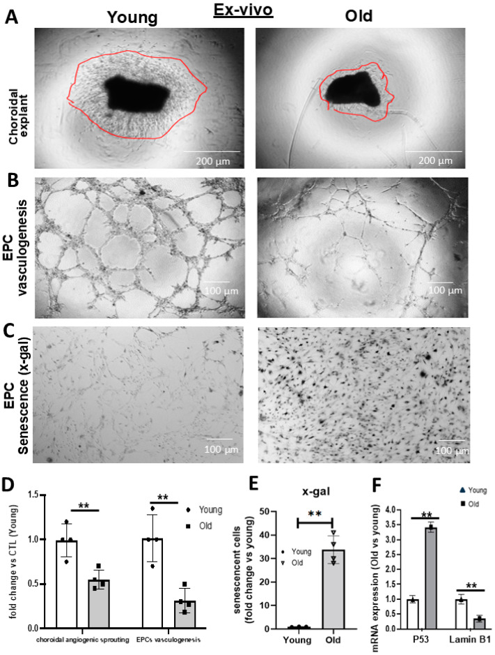
Background: Choroidal involution is a common feature of age-related ischemic retinopathies such as age-related macular degeneration (AMD). It is now well recognized that endothelial progenitor cells (EPCs) are essential to endothelial repair processes and in maintaining vascular integrity. However, the contribution of EPCs and the role of senescence in age-related choroidal vascular degeneration remain to be investigated. In this study, we compared the senescent phenotype of EPCs in the choroid and performed whole-genome profiling of EPCs derived from young versus old rats. Methods and Results: We isolated and compared the retinas of young (6-weeks-old) and old (16-18-month-old) rats. The thickness of the choroid and outer nuclear layer (ONL), along with local quantification of CD34+ EPCs, was performed. Compared to young rats, older rats displayed a significant reduction in choroidal and ONL thickness associated with markedly fewer choroid-localized EPCs; this was attested by lower expression of several EPC markers (CXCR4, CD34, CD117, CD133, and KLF-2). Choroid and choroid-localized EPCs displayed abundant senescence as revealed by increased β-gal and P53 expression and decreased Lamin-B1 (immunostaining and RT-qPCR). Concordantly, choroidal cells and EPCs isolated from older rats were unable to form vascular networks ex vivo. To better understand the potential mechanisms associated with the dysfunctional EPCs linked to age-related choroidal involution, we performed whole-genome profiling (mRNA and miRNA) of EPCs derived from old and young rats using next-generation sequencing (NGS); 802 genes were significantly modulated in old vs. young EPCs, corresponding to ~2% of total genes expressed. Using a bioinformatic algorithm, the KEGG pathways suggested that these genes participate in the modulation of several key signaling processes including inflammation, G protein-coupled receptors, and hematopoietic cell lineages. Moreover, we identified 13 miRNAs involved in the regulation of immune system processes, cell cycle arrest and senescence, which are significantly modulated in EPCs from old rats compared to young ones. Conclusions: Our results suggest that age-related choroidal involution is associated with fewer EPCs, albeit displaying a senescence-like phenotype. One would be tempted to propose that biological modification of native EPCs (such as with senolytic agents) could potentially provide a new strategy to preserve the vascular integrity of the aged choroid, and evade progression to degenerative maculopathies.

Keywords: aging; choroidal involution; endothelial progenitor cell (EPC); senescence; vascular network.


Conflict of interest statement

The authors declare that the research was conducted in the absence of any commercial or financial relationships that could be construed as potential conflicts of interest.

Figures

Figure 1
Figure 1
Choroidal thickness and EPC number in young compared to old choroids. (A,C) Representative images (A) and quantitative analyses (C) of choroidal (CHR) and photoreceptor (OPL) thickness in retinal cross-sections stained for vessels (isolectin-positive, green) and DAPI in old vs. young rats. (B,D) EPCs (CD34+ cells) co-localized in choroidal vessels in old vs. young rats. For the choroid of old animals in the inset in (B), the magnification is 10-fold greater than that in young animals to make it possible to compare CD34 expression over the same choroidal vessel area. Data are mean ± SEM. ** p < 0.01 or *** p < 0.001 vs. young rats. n = 5.
Figure 2
Figure 2
Comparison ex vivo of choroidal angiogenic sprouting, EPC vasculogenic capacity and senescence level in young vs. old rats. (A,B,D) Representative images and quantitative analysis of the angiogenic capacity of isolated choroidal explants (A,D) at day 5 and (B) EPCs at 6 h, derived from young vs. old rats, as assessed by the Matrigel assay. (C,E) EPC senescence level assessed by x-gal assay. (F) qRT-PCR analysis of P53 and Lamin B1 expression. Data were mean ± SEM. ** p < 0.01 vs. young rats. n = 5.
Figure 3
Figure 3
Senescence level in the choroid and in choroidal EPCs in young compared to old rats. (A,B) Representative images and quantification analysis of β-gal expression and LaminB1 in the choroidal vessels of young vs. old rats. (C) Representative image of p53 expression localized on choroidal vessels. (D) Representative image of p53 expression in EPCs co-localized in choroidal vessels. The white vertical bar in panel A identifies the thickness of the choroid (CHR). The image for the (B) panel in old animals is magnified 10-fold compared to images for young animals to compare the expression of p53 per same vessel area. The fluorescence intensity was measured for the stained markers (P53 and laminB1) per total choroid area using ImageJ. The histogram insets refer to expression profiles normalized to the choroids of young rats. Data were mean ± SEM. ** p < 0.01 vs. young rats. n = 5.
Figure 4
Figure 4
Global NGS analysis showing modulated genes and the associated altered pathways in EPCs derived from old rats. (A,B) Global evaluation of significant (p < 0.05) modulated gene (mRNA) expression levels (arbitrary cut-off levels set at a 2-log-fold change) in EPCs derived from old rats compared to young rats (A) and graphical representation in terms of the percentage of the distribution of the modulated genes in the whole genome (B). (C) Bioinformatic analysis identifying pathway enrichment (ranking by p value probability) by the modulation of the 802 mRNAs identified; the table on right side ranks pathways based on a combined score (of z-value deviation × the log of the p value score). The NGS data set represents the mRNA expression level in EPCs extracted in n = 3 rats per group.
Figure 5
Figure 5
NGS analysis of the effect of aging on the expression level of miRNAs in EPCs and the associated pathway altered. (A) Global evaluation of significant (p < 0.05) modulated miRNAs (arbitrary cut-off levels set at a 2-log-fold change) in EPCs derived from old rats compared to young rats and a graphical representation in percentage terms of the miRNA distribution. (B) Heat map showing the identity and the logFC of the 13 miRNAs identified as significantly modulated. (C) KEGG analysis to identify predicted pathways altered (ranked by the p value of probability) by the simultaneous modulation of the five modulated miRNAs identified to be also expressed in humans. Blue box represent the pathway that potentially affect EPCs function. The NGS data set represents miRNA expression levels in EPCs extracted in n = 3 rats per group.

Similar articles

Cited by

References

    1. Tisi A., Feligioni M., Passacantando M., Ciancaglini M., Maccarone R. The Impact of Oxidative Stress on Blood-Retinal Barrier Physiology in Age-Related Macular Degeneration. Cells. 2021;10:64. doi: 10.3390/cells10010064. - DOI - PMC - PubMed
    1. Harris A., Bingaman D., Ciulla T.A., Martin B. Chapter 5—Retinal and Choroidal Blood Flow in Health and Disease. In: Ryan S.J., Hinton D.R., Schachat A.P., Wilkinson C.P., editors. Retina. 4th ed. Mosby; Edinburgh, UK: 2006. pp. 83–102. - DOI
    1. Desjarlais M., Rivera J.C., Lahaie I., Cagnone G., Wirt M., Omri S., Chemtob S. MicroRNA expression profile in retina and choroid in oxygen-induced retinopathy model. PLoS ONE. 2019;14:e0218282. doi: 10.1371/journal.pone.0218282. - DOI - PMC - PubMed
    1. Fragiotta S., Scuderi L., Iodice C.M., Rullo D., Di Pippo M., Maugliani E., Abdolrahimzadeh S. Choroidal Vasculature Changes in Age-Related Macular Degeneration: From a Molecular to a Clinical Perspective. Int. J. Mol. Sci. 2022;23:12010. doi: 10.3390/ijms231912010. - DOI - PMC - PubMed
    1. Zhou T.E., Rivera J.C., Bhosle V.K., Lahaie I., Shao Z., Tahiri H., Zhu T., Polosa A., Dorfman A., Beaudry-Richard A., et al. Choroidal Involution Is Associated with a Progressive Degeneration of the Outer Retinal Function in a Model of Retinopathy of Prematurity: Early Role for IL-1β. Am. J. Pathol. 2016;186:3100–3116. doi: 10.1016/j.ajpath.2016.08.004. - DOI - PubMed

LinkOut - more resources