. 2024 Dec 14;12(12):2847.
doi: 10.3390/biomedicines12122847.

Fucoidan Attenuates Cardiac Remodeling by Inhibiting Galectin-3 Secretion, Fibrosis, and Inflammation in a Mouse Model of Pressure Overload

Affiliations

Fucoidan Attenuates Cardiac Remodeling by Inhibiting Galectin-3 Secretion, Fibrosis, and Inflammation in a Mouse Model of Pressure Overload

Wen-Rui Hao et al. Biomedicines. .

Abstract

Background/objectives: Fucoidan, a sulfated polysaccharide derived from marine algae, is known for its antioxidant and immunomodulatory properties. Galectin-3 (Gal-3), a protein associated with cardiovascular fibrosis, has been identified as a potential therapeutic target in cardiac remodeling. This study aimed to evaluate whether fucoidan could inhibit Gal-3 activity and mitigate cardiac remodeling in a mouse model of pressure overload-induced cardiac hypertrophy.

Methods: To test this hypothesis, we used transverse aortic constriction (TAC) surgery to induce pressure overload in normotensive mice, replicating the pathological features of cardiac hypertrophy. Mice were treated with fucoidan at a dose of 1.5 or 7.5 mg/kg/day. In vivo assessments of cardiac function, fibrosis, inflammation, and Gal-3 expression were performed.

Results: Pressure overload led to significant upregulation of serum Gal-3 levels, increased cardiac collagen deposition, and elevated markers of fibrosis and inflammation. In mice treated with fucoidan, these effects were significantly attenuated. Fucoidan treatment prevented the upregulation of Gal-3, reduced collagen deposition, and decreased inflammatory cell infiltration, suggesting an inhibition of both fibrosis and inflammation.

Conclusions: Fucoidan effectively mitigated the adverse effects of pressure overload in this mouse model, including reduced Gal-3 expression, fibrosis, and inflammation. These findings suggest that fucoidan holds promise as a therapeutic agent for preventing or delaying cardiac remodeling and associated complications, such as fibrosis and inflammation, in pressure overload-induced cardiac hypertrophy. Further research is needed to explore the underlying mechanisms and clinical applicability of fucoidan in cardiac disease.

Keywords: fibrosis; fucoidan; galectin-3; pressure overload.


Conflict of interest statement

The authors declare no conflicts of interest.

Figures

Figure 1
Figure 1
Timeline of fucoidan (FO) administration and tissue collection in TAC-induced cardiac remodeling model. Male C57BL/6J mice (8–10 weeks, 23.5–27.5 g) underwent TAC surgery to induce cardiac hypertrophy and fibrosis or sham surgery as a control. Mice received FO by oral gavage starting one day before TAC surgery and continuing daily until tissue collection at 3 weeks post-surgery. FO was administered in two doses: low-dose fucoidan (FOL; 60 mg/kg/day, equivalent to 1.5 mg/day for a 25 g mouse) and high-dose fucoidan (FOH; 300 mg/kg/day, equivalent to 7.5 mg/day for a 25 g mouse) for 14 days. The arrow represents the timeline of the study, indicating the duration and sequence of fucoidan administration and tissue collection. At the end of the experiment, tissues were harvested to assess the effects of FO on cardiac remodeling.
Figure 2
Figure 2
Effects of Fucoidan on whole heart and left ventricular tissue weights, as well as on body weight under varying experimental conditions. (A) Heart images from each experimental group visually depict the impact of Fucoidan treatment. (B) The heart weight-to-body weight ratio demonstrates significant variations across groups, with TAC surgery inducing notable increases in both metrics. Fucoidan treatment, however, reduced these ratios compared to the TAC-induced cardiac hypertrophy group. Experimental groups included the sham-operated control (sham), TAC-induced cardiac hypertrophy group (TAC), TAC group treated with 1.5 mg/kg/day Fucoidan (FOL), and TAC group treated with 7.5 mg/kg/day Fucoidan (FOH). Data are presented as means ± standard deviation (SD) with n = 5. *** p < 0.001 vs. sham; and # p < 0.05 and ## p < 0.01 vs. TAC.
Figure 3
Figure 3
Effects of Fucoidan on cardiac function in a mouse model of transverse aortic constriction (TAC)-induced cardiac hypertrophy. (A) Representative echocardiographic images from each experimental group illustrate changes in cardiac function. (B) Analysis of diastolic wall strain (DWS) across groups reveals significant differences. The TAC-induced cardiac hypertrophy group exhibited a reduced ejection fraction compared to the sham-operated control, while Fucoidan treatment partially restored the ejection fraction in both treatment groups. DWS, calculated from posterior wall thickness (PWT) measurements as DWS = [PWT(systole) − PWT(diastole)]/PWT(systole), serves as a marker for left ventricular (LV) diastolic stiffness. The experimental groups included sham-operated controls (sham), the TAC-induced hypertrophy group (TAC), and TAC groups treated with 1.5 mg/kg/day (FOL) or 7.5 mg/kg/day Fucoidan (FOH). Data are expressed as means ± standard deviation (SD), with n = 5. Statistical significance is indicated by and *** p < 0.001 vs. sham, and ## p < 0.01, and ### p < 0.001 vs. TAC.
Figure 4
Figure 4
Effects of Fucoidan on histopathological changes in mouse heart tissue. (A) Representative images of Masson staining (top panels) and Sirius red staining (bottom panels) of left ventricular tissue from each group are shown at 200× magnification, with scale bars indicating 1000 μm. (B,C) Quantitative analyses display the area percentage of myocardial damage and collagen deposition (n = 5), with data presented as mean ± standard deviation (SD). Statistical significance is noted as *** p < 0.001, compared to the control sham group, and ### p < 0.001, compared to the TAC group.
Figure 5
Figure 5
Effect of fucoidan on serum Galectin-3 (Gal-3) levels in mice following transverse aortic constriction (TAC). Serum Gal-3 levels were measured by enzyme-linked immunosorbent assay (ELISA) at day 22 post-TAC. The TAC group exhibited a significant increase in Gal-3 levels compared to the sham group, indicating fibrosis due to pressure overload. Treatment groups receiving fucoidan (FOL and FOH) showed a marked reduction in Gal-3 levels compared to the TAC group, suggesting fucoidan’s potential to mitigate fibrosis-related signaling. Data are expressed as mean ± standard deviation (SD) (n = 5). Statistical significance is indicated as *** p < 0.001 vs. sham, and ### p < 0.001 vs. TAC.
Figure 6
Figure 6
Fucoidan modulates key inflammatory and fibrotic signaling pathways, highlighting its potential as a therapeutic candidate for cardiovascular and fibrotic diseases. This study evaluates the effects of fucoidan on pro-inflammatory and fibrotic signaling pathways using Western blot analysis to detect specific markers associated with inflammation and fibrosis. The markers analyzed include Galectin-3 (Gal-3), CD68, connective tissue growth factor (CTGF), discoidin domain receptor 2 (DDR2), alpha-smooth muscle actin (α-SMA), CD44, and collagen I, providing insights into fucoidan’s regulatory effects on these pathways. (A) Protein levels of CD68, CTGF, DDR2, α-SMA, CD44, and collagen I in each group on day 22 are shown. (BH) Quantification of protein bands for CD68, CTGF, DDR2, α-SMA, CD44, and collagen I, normalized to GAPDH as the internal control (n = 3). Data are presented as mean ± standard deviation (SD). Statistical significance is indicated as follows: * p < 0.05 vs. sham group; ** p < 0.01 vs. sham group; # p < 0.05 vs. TAC group; ## p < 0.01 vs. TAC group.
Figure 7
Figure 7
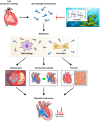
Summary of fucoidan’s effects on cardiac remodeling and inflammation in pressure overload-induced cardiac hypertrophy. This figure illustrates the protective effects of fucoidan against cardiac remodeling, fibrosis, and inflammation in a mouse model of pressure overload-induced cardiac hypertrophy generated by transverse aortic constriction (TAC). TAC surgery was used to induce pressure overload, resulting in pathological cardiac hypertrophy. Following aortic constriction, there was a significant increase in cardiac Galectin-3 (Gal-3) expression, a protein closely associated with fibrosis and inflammation. Elevated Gal-3 levels correlated with increased collagen deposition and upregulation of fibrotic mediators, indicative of cardiac fibrosis. In addition, TAC-induced pressure overload led to increased levels of inflammatory markers and a marked infiltration of inflammatory cells into cardiac tissue, reflecting an inflammatory response associated with cardiac hypertrophy. Fucoidan treatment effectively mitigated the adverse effects of pressure overload. Fucoidan administration prevented the upregulation of Gal-3, resulting in decreased collagen deposition and downregulation of fibrotic mediators, indicating attenuation of cardiac fibrosis. Additionally, fucoidan significantly reduced inflammatory cell infiltration and lowered inflammatory cytokine levels in TAC mice, highlighting its anti-inflammatory effects. These findings suggest that fucoidan can delay or prevent the progression of cardiac remodeling in pressure overload by inhibiting Gal-3, reducing fibrosis, and decreasing inflammation. Fucoidan shows promise as a therapeutic agent for mitigating Gal-3 overexpression and associated cardiac complications. The arrow indicates the direction of the observed effect, while the red hammer symbolizes the inhibitory effect.

References

    1. Aghajanian H., Kimura T., Rurik J.G., Hancock A.S., Leibowitz M.S., Li L., Scholler J., Monslow J., Lo A., Han W., et al. Targeting cardiac fibrosis with engineered T cells. Nature. 2019;573:430–433. doi: 10.1038/s41586-019-1546-z. - DOI - PMC - PubMed
    1. Beslika E., Leite-Moreira A., De Windt L.J., da Costa Martins P.A. Large animal models of pressure overload-induced cardiac left ventricular hypertrophy to study remodelling of the human heart with aortic stenosis. Cardiovasc. Res. 2024;120:461–475. doi: 10.1093/cvr/cvae045. - DOI - PMC - PubMed
    1. Hao W.R., Cheng C.H., Liu J.C., Chen H.Y., Chen J.J., Cheng T.H. Understanding Galectin-3’s Role in Diastolic Dysfunction: A Contemporary Perspective. Life. 2024;14:906. doi: 10.3390/life14070906. - DOI - PMC - PubMed
    1. An L., Chang G., Zhang L., Wang P., Gao W., Li X. Pectin: Health-promoting properties as a natural galectin-3 inhibitor. Glycoconj. J. 2024;41:93–118. doi: 10.1007/s10719-024-10152-z. - DOI - PubMed
    1. Martínez-Martínez E., Calvier L., Fernández-Celis A., Rousseau E., Jurado-López R., Rossoni L.V., Jaisser F., Zannad F., Rossignol P., Cachofeiro V., et al. Galectin-3 blockade inhibits cardiac inflammation and fibrosis in experimental hyperaldosteronism and hypertension. Hypertension. 2015;66:767–775. doi: 10.1161/HYPERTENSIONAHA.115.05876. - DOI - PubMed

LinkOut - more resources