Review
. 2024 Dec 13;13(24):7605.
doi: 10.3390/jcm13247605.

Three-Dimensional Ultrasound for Physical and Virtual Fetal Heart Models: Current Status and Future Perspectives

Affiliations
Review

Three-Dimensional Ultrasound for Physical and Virtual Fetal Heart Models: Current Status and Future Perspectives

Nathalie Jeanne Bravo-Valenzuela et al. J Clin Med. .

Abstract

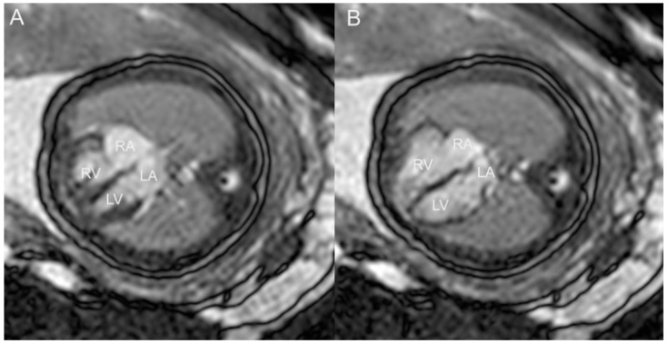
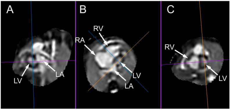
Congenital heart defects (CHDs) are the most common congenital defect, occurring in approximately 1 in 100 live births and being a leading cause of perinatal morbidity and mortality. Of note, approximately 25% of these defects are classified as critical, requiring immediate postnatal care by pediatric cardiology and neonatal cardiac surgery teams. Consequently, early and accurate diagnosis of CHD is key to proper prenatal and postnatal monitoring in a tertiary care setting. In this scenario, fetal echocardiography is considered the gold standard imaging ultrasound method for the diagnosis of CHD. However, the availability of this examination in clinical practice remains limited due to the need for a qualified specialist in pediatric cardiology. Moreover, in light of the relatively low prevalence of CHD among at-risk populations (approximately 10%), ultrasound cardiac screening for potential cardiac anomalies during routine second-trimester obstetric ultrasound scans represents a pivotal aspect of diagnosing CHD. In order to maximize the accuracy of CHD diagnoses, the views of the ventricular outflow tract and the superior mediastinum were added to the four-chamber view of the fetal heart for routine ultrasound screening according to international guidelines. In this context, four-dimensional spatio-temporal image correlation software (STIC) was developed in the early 2000s. Some of the advantages of STIC in fetal cardiac evaluation include the enrichment of anatomical details of fetal cardiac images in the absence of the pregnant woman and the ability to send volumes for analysis by an expert in fetal cardiology by an internet link. Sequentially, new technologies have been developed, such as fetal intelligent navigation echocardiography (FINE), also known as "5D heart", in which the nine fetal cardiac views recommended during a fetal echocardiogram are automatically generated from the acquisition of a cardiac volume. Furthermore, artificial intelligence (AI) has recently emerged as a promising technological innovation, offering the potential to warn of possible cardiac anomalies and thus increase the ability of non-cardiology specialists to diagnose CHD. In the early 2010s, the advent of 3D reconstruction software combined with high-definition printers enabled the virtual and 3D physical reconstruction of the fetal heart. The 3D physical models may improve parental counseling of fetal CHD, maternal-fetal interaction in cases of blind pregnant women, and interactive discussions among multidisciplinary health teams. In addition, the 3D physical and virtual models can be an useful tool for teaching cardiovascular anatomy and to optimize surgical planning, enabling simulation rooms for surgical procedures. Therefore, in this review, the authors discuss advanced image technologies that may optimize prenatal diagnoses of CHDs.

Keywords: artificial intelligence; congenital heart disease; fetal heart; fetal intelligent navigation echocardiography; spatiotemporal image correlation; three-dimensional ultrasound; ultrasonography.


Conflict of interest statement

The authors declare no conflicts of interest.

Figures

Figure 1
Figure 1
Measurements of interventricular septum volume (IVS) using 3D ultrasound with STIC and virtual organ computer-aided analysis (VOCAL) in a fetus from a diabetic mother at 25 weeks of gestation. IVS volume = 0.144 cm3.
Figure 2
Figure 2
Left ventricle diastolic volume using STIC with virtual organ computer-aided analysis (VOCAL) in a fetus at 30 weeks of gestation. LV volume = 1.3 cm3.
Figure 3
Figure 3
Evaluation of the tricuspid annular movement using fetal STIC-M (5.4 mm). TAPSE: tricuspid annular plane systolic excursion; RV: right ventricle.
Figure 4
Figure 4
Three-dimensional ultrasound with STIC: (A) HDlive mode, providing a reconstruction of the left ventricular outflow tract in a case of transposition of the great arteries and (B) with color Doppler in a first-trimester fetus with tetralogy of Fallot. Observe the pulmonary artery (P) arising from the left ventricle (LV) in image (A) and the overriding of the aorta (A) in image (B). RV: right ventricle; LV: left ventricle; VSD: ventricular septal defect; IVS: ventricular septum.
Figure 5
Figure 5
Tomographic ultrasound imaging (TUI) in the rendering mode enables the visualization of sequential axial planes in the case of inlet ventricular septal defect (VSD) (yellow arrows).
Figure 6
Figure 6
STIC with HDlive Silhouette mode in a case of coarctation of aorta. Note the discrepancy of the great arteries due to the small aorta. AO: aorta; PA: pulmonary artery; VC: superior vena cava.
Figure 7
Figure 7
(A) Three-dimensional ultrasound with Surface Realistic Vue (SRV) imaging in a case of partial anomalous pulmonary vein return with a ventricular septal defect (VSD). Note that 2 of the pulmonary veins return to the right atrium (red arrows). Virtual light source position, 10 o’clock. (B) STIC with color Doppler of a case of total anomalous pulmonary vein return (infradiaphragmatic type). The right (RPV) and left pulmonary veins (LPVs) drain (white arrows) into a collecting vein (COL) and subsequently into a vertical vein (VV), which achieves the right atrium (RA) via the inferior vena cava (IVC). LV: left ventricle; LA: left atrium; RA: right atrium; RV: right ventricle; ** VSD: ventricular septal defect; PV: pulmonary vein; T: tricuspid valve; M: mitral valve.
Figure 8
Figure 8
Three-dimensional ultrasound with STIC and HDlive mode in a case of left heterotaxy. Observe that the venous vessel (hemiazygos) is located posteriorly (near to the fetal spine) to the arterial vessel (aorta) at the upper abdomen view. Ao; aorta; Hz: hemiazygos vein; L: fetal left side; R: fetal right side.
Figure 9
Figure 9
Extra-hepatic form of agenesis of ductus venosus using three-dimensional ultrasound with STIC. Note the high-resolution color Doppler showing the absence of flow through the DV (red arrow). In this case, the umbilical vein drains into the RA via the inferior vena cava. IVC: inferior vena cava; RA: right atrium.
Figure 10
Figure 10
Three-dimensional ultrasound with STIC enabling the reconstruction of the ventricular outflow tracts in a case of double-outlet right vetricle (“Taussig-Bing” anomaly). Note the great arteries arising from the right ventricle (RV) in a parallel relationship. Ao: aorta; PA: pulmonary artery.
Figure 11
Figure 11
Tomographic ultrasound imaging (TUI) in the rendering mode in a case of tetralogy of Fallot and in (B) double outflow of the right ventricle (DORV). The right ventricle hypertrophy (yellow arrows) could be observed using this technology (A). Note the great arteries in a parallel relationship (red arrows) in a fetus with Taussig–Bing DORV using color Doppler (B). DORV: double outflow of right ventricle; Ao; aorta; P: pulmonary artery.
Figure 12
Figure 12
The reconstruction of the ventricular outflow tracts in a case of transposition of the great arteries (TGA) using STIC with color Doppler (A) and HDlive Silhouette. In image (A), it is evident that the aorta (Ao) arises from the right ventricle (RV). In image (B), the pulmonary artery (PA) is unequivocally identified as originating from the left ventricle (LV). The two arteries are observed to be in a parallel relationship (red arrows), with the aorta located anteriorly to the PA.
Figure 13
Figure 13
Three-dimensional ultrasound with STIC in the rendering mode: the measurement of the area of the foramen ovale (FO) was obtained from the four-chamber view of the fetal heart in which the ROI (green line) is the flap of the FO. ROI: region of interest.
Figure 14
Figure 14
Three-dimensional with STIC in the rendering mode (A) and HDlive mode (B) of a fetus with Ebstein’s anomaly at 30 weeks of gestation. RA: right atrium; T: tricuspid valve; RV: right ventricle; LA: left atrium; M: mitral valve LV: left ventricle.
Figure 15
Figure 15
(A) Reconstruction of the aortic arch using STIC with the inversion mode in a case of coarctation of the aorta. Observe the narrowing of the aortic isthmus (yellow arrow). (B) Aortic and duct arch imaging in a fetus with a normal heart. (B) Sagittal view of a fetus with a normal heart showing the aortic and ductal arches using LumiFlow. (C) First-trimester imaging using HDFlow in a fetus with a right aortic arch (red arrow) and vascular ring (observe the vessels around the trachea). Ao: aorta; BCT: brachiocephalic trunk; LCC: left common carotid; LSCA: left subclavian artery; P: pulmonary artery; DA: ductus arteriosus; Tr: trachea; R: right side; L: left side.
Figure 16
Figure 16
Large mass (**) in the ventricular septum and both ventricles, mainly in the left ventricle, in a case of rhabdomyomas with a reduction in the size of the masses after prenatal therapy with sirolimus. LV: left ventricle; LA: left atrium; RA: right atrium; RV: right ventricle; T: tricuspid valve.
Figure 17
Figure 17
STIC-M enabling the measurement of mitral annular plane systolic excursion (MAPSE) (5.4 mm). LV: left ventricle.
Figure 18
Figure 18
Three-dimensional reconstruction of the left ventricle (LV) using STIC with virtual organ computer-aided analysis (VOCAL) in a fetus at 22 weeks of gestation.
Figure 19
Figure 19
FINE navigation (known as “5D heart”) in (A) a case of a malalignment type of ventricular septal defect (***, yellow arrows) and in (B) a case of complete atrioventricular septal defect (AVSD). In case (A), observe the overriding of the aorta (Ao). In case (B), observe that the four-chamber, the five-chamber, and LV outflow tract (LVOT) views (yellow arrows) draw attention to this diagnosis. *** Common AV valve; VSD: ventricular septal defect; ASD: primum atrial septal defect; GN: LVOT with a “goose neck” shape.
Figure 20
Figure 20
Automatic measurement of the fetal the cardiac axis (40.3º) using artificial intelligence (“Learning Machine”) in a normal heart using fetal intelligent navigation echocardiography (FINE), also known as “5D Heart”. LV: left ventricle; LA: left atrium; RA: right atrium; RV: right ventricle; A or Ao: aorta; P or PA: pulmonary artery; S: superior vena cava; IVC: inferior vena cava; Desc: descending; Trans: transverse.
Figure 21
Figure 21
First-trimester measurement of the cardiac axis (45°) of a normal fetus (yellow arrow). L: left side; R:right side: Ao: aorta; S: spine.
Figure 22
Figure 22
Three-dimensional physical model of a fetus with transposition of the great arteries (TGA). RV: right ventricle; Ao: aorta; LV: left ventricle; P: pulmonary artery.
Figure 23
Figure 23
Three-dimensional virtual model of fetal heart in a fetus with transposition of the great arteries (TGA) (A) and in a fetus with Ebstein’s anomaly (B). RA: right atrium; RV: right ventricle; LA: left atrium; T: tricuspid valve; M: mitral valve; LV: left ventricle; Ao: aorta; P: pulmonary artery.
Figure 24
Figure 24
Following the acquisition of images of the fetal heart with tetralogy of Fallot from 3D ultrasound (heart volumes) using tools from Slicer 3D software (Birmingham, UK), the cardiac structures were segmented, with each cavity identified by a different color (right and left atrium, right and left ventricles, aorta, pulmonary artery, vena cava and pulmonary veins). Thereafter, a raw file format was generated. Based on the 3D data, physical 3D models of the fetal heart were printed using a 3D printer. Ao: aorta; LA: left atrium; P: pulmonary artery; RA: right atrium; LV: left ventricle; RV: right ventricle; VSD: ventricular septal defect.
Figure 25
Figure 25
(A) Fetal cardiac MRI (fCMR) performed at 32 weeks and 5 days. Images were obtained at 1.5 T using a balanced turbo field echo (BTFE) sequence, gated with an MRI-compatible Doppler ultrasound (DUS) device (North Medical, Hamburg, Germany). Four-chamber view in systole (A) and diastole (B). LA: left atrium; LV: left ventricle; RA: right atrium; LA: left atrium.
Figure 26
Figure 26
Multiplanar display images of a case of hypoplastic left heart syndrome examined at 32 weeks and 3 days. The images were acquired using a balanced turbo field echo (BTFE) sequence at 1.5 T. kt-sense acceleration was used during acquisition. The images were postprocessed using a super-resolution pipeline, resulting in an isovoxel 3D volume dataset. (A) Sagittal two-chamber view. (B) Four-chamber view. (C) Coronal short-axis view through the ventricles. LA: left atrium; LV: left ventricle; RA: right atrium; RV: right ventricle.

Similar articles

References

    1. Liu Y., Chen S., Zühlke L., Black G.C., Choy M.K., Li N., Keavney B.D. Global birth prevalence of congenital heart defects 1970-2017: Updated systematic review and meta-analysis of 260 studies. Int. J. Epidemiol. 2019;48:455–463. doi: 10.1093/ije/dyz009. - DOI - PMC - PubMed
    1. Bouma B.J., Mulder B.J. Changing Landscape of Congenital Heart Disease. Circ. Res. 2017;120:908–922. doi: 10.1161/CIRCRESAHA.116.309302. - DOI - PubMed
    1. Hoffman J.I.E. The global burden of congenital heart disease. Cardiovasc. J. Afr. 2013;24:141–145. doi: 10.5830/CVJA-2013-028. - DOI - PMC - PubMed
    1. Khasawneh W., Hakim F., Abu Ras O., Hejazi Y., Abu-Aqoulah A. Incidence and patterns of congenital heart disease among Jordanian infants, a cohort study from a university tertiary center. Front. Pediatr. 2020;8:219. doi: 10.3389/fped.2020.00219. - DOI - PMC - PubMed
    1. Meller C.H., Grinenco S., Aiello H., Córdoba A., Sáenz-Tejeira M.M., Marantz P., Otaño L. Congenital heart disease, prenatal diagnosis and management. Arch. Argent. Pediatr. 2020;118:e149–e161. - PubMed

LinkOut - more resources