Review
. 2024 Dec 20;13(24):7790.
doi: 10.3390/jcm13247790.

Diagnostic and Therapeutic Approach in Pediatric Pulmonary Abscess: Two Cases and Literature Review

Affiliations
Review

Diagnostic and Therapeutic Approach in Pediatric Pulmonary Abscess: Two Cases and Literature Review

Mariana Costin et al. J Clin Med. .

Abstract

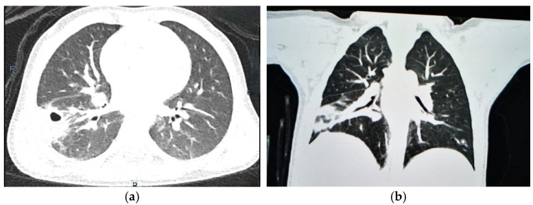
Pulmonary abscess is a rare but serious condition in pediatric patients, caused by severe pulmonary infection that leads to tissue destruction and necrosis. It can be classified as primary or secondary depending on the cause. Establishing an etiology in pediatric pulmonary abscesses is challenging, underscoring the essential role of advanced imaging techniques, such as computed tomography, in achieving an accurate diagnosis and differentiating among various conditions that may mimic lung abscess. While conservative management with antibiotics is the first line of treatment, some cases may progress and require surgical intervention. We present two clinical cases of pediatric lung abscesses, emphasizing the importance of timely intervention, accompanied by a brief review of current knowledge that highlights key clinical features, diagnostic challenges, and therapeutic approaches in pediatric lung abscess.

Keywords: antibiotics; children; empyema; pulmonary abscess; thoracoscopy; video-assisted thoracic surgery (VATS).


Conflict of interest statement

The authors declare no conflict of interest.

Figures

Figure 1
Figure 1
Two-year old male child with pulmonary abscess: oval mass with air–fluid level and blurred margins in right lower lobe; (a) lateral chest X-ray; (b) posteroanterior chest X-ray.
Figure 2
Figure 2
Two-year old male child with pulmonary abscess: thoracic ultrasonography showing (a) round–oval lesion with heterogeneous anechoic content, with no adjacent pleural effusion; (b) absent intrinsic vascular signal, but peripheral parietal vascular tract is present, suggestive of underlying congenital pulmonary airway malformation.
Figure 3
Figure 3
Two-year-old male child with pulmonary abscess: CT scan revealed large mass in right lower lobe, measuring approximately 53 mm × 47 mm × 50 mm, containing heterogeneous contents enclosed by thick walls. Two additional cystic lesions, measuring 13 mm and 7 mm, respectively, observed adjacent to mass without clear demarcation, indicative of multiloculated cystic lesion centered in right lower lobe; (a) transverse plane; (b) coronal plane.
Figure 4
Figure 4
CT scan of 2-year-old male child with pulmonary abscess, demonstrating marked reduction in hydro-aeric cavity in right lower lobe, measuring 22 mm × 22 mm, without associated cystic lesions; (a) transverse plane; (b) coronal plane.
Figure 5
Figure 5
Normal chest X-ray of 2-year-old male child with pulmonary abscess, 3 months post-treatment.
Figure 6
Figure 6
Seventeen-year-old male adolescent with pulmonary abscess: chest X-ray demonstrating oval-shaped, well-circumscribed opacity occupying two-thirds of right hemithorax, characterized by thick walls and containing air–fluid level; satellite lymphadenopathies observed in right hilum.
Figure 7
Figure 7
Seventeen-year-old male adolescent with pulmonary abscess: CT scan (transverse plane) with contrast performed, which describes oval mass in inferior right lobe, measuring circa 10 cm × 7 cm × 12 cm, with some round–oval parts containing air–fluid levels, circumscribed by walls and septa with varying thickness; pleural effusion of maximum thickness 10 mm.
Figure 8
Figure 8
Seventeen-year-old male adolescent with pulmonary abscess: enlargement of oval-shaped mass, measuring approximately 15 cm × 9 cm × 12 cm, characterized by thick walls and containing two air–fluid levels observed in upper third of mass, with increased radiopaque component noted; (a) posteroanterior chest X-ray; (b) lateral chest X-ray.
Figure 9
Figure 9
Seventeen-year-old male adolescent with pulmonary abscess: (a) post-operative pneumothorax requiring pleural drainage, initially with poor lung expansion; (b) after three days, right lung expanded adequately.
Figure 10
Figure 10
Seventeen-year-old male adolescent with pulmonary abscess—histopathologic results: (a) 4× magnification objective: disorganized architecture with alveoli and bronchioles not visible, replaced by fibroblastic reaction and mixed inflammatory infiltrate, indicative of abscess formation; (b) 10× magnification objective: mixed inflammatory infiltrate (lymphoplasmacytes, neutrophils, eosinophils, and macrophages) in interalveolar septa and bronchial epithelium; (c) 20× magnification objective: alveoli filled with macrophages and thickened interalveolar septa with inflammatory infiltrate consisting of lymphocytes, plasma cells, neutrophils, and eosinophils.

Similar articles

Cited by

References

    1. Patradoon-Ho P., Fitzgerald D.A. Lung abscess in children. Paediatr. Respir. Rev. 2007;8:77–84. doi: 10.1016/j.prrv.2006.10.002. - DOI - PubMed
    1. De Benedictis F.M., Kerem E., Chang A.B., Colin A.A., Zar H.J., Bush A. Complicated pneumonia in children. Lancet. 2020;396:786–798. doi: 10.1016/S0140-6736(20)31550-6. - DOI - PubMed
    1. Yousef L., Yousef A., Al-Shamrani A. Lung Abscess Case Series and Review of the Literature. Children. 2022;9:1047. doi: 10.3390/children9071047. - DOI - PMC - PubMed
    1. Kuhajda I., Zarogoulidis K., Tsirgogianni K., Tsavlis D., Kioumis I., Kosmidis C., Tsakiridis K., Mpakas A., Zarogoulidis P., Zissimopoulos A., et al. Lung abscess-etiology, diagnostic and treatment options. Ann. Transl. Med. 2015;3:183. - PMC - PubMed
    1. Roik D., Mosior T., Sopyło B., Małdyk J., Brzewski M. Abscess mimicking lung metastasis in a 10-year-old boy—Case report. Pol. J. Radiol. 2010;75:55–57. - PMC - PubMed

LinkOut - more resources