Review
. 2024 Dec 10;25(24):13238.
doi: 10.3390/ijms252413238.

The Causal Role of Ectopic Fat Deposition in the Pathogenesis of Metabolic Syndrome

Affiliations
Review

The Causal Role of Ectopic Fat Deposition in the Pathogenesis of Metabolic Syndrome

Joseph A M J L Janssen. Int J Mol Sci. .

Abstract

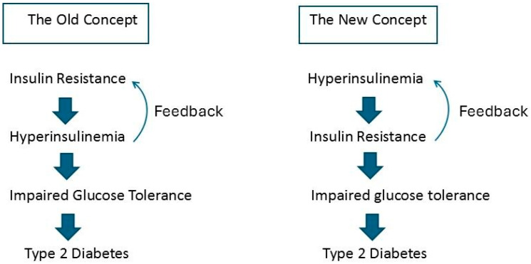
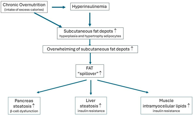
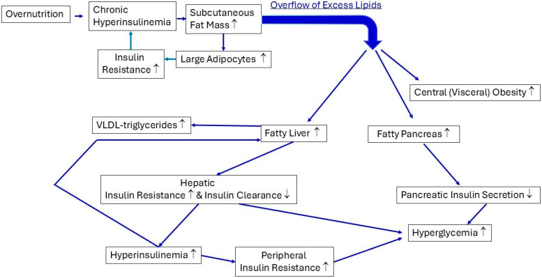
Consuming a "modern" Western diet and overnutrition may increase insulin secretion. Additionally, nutrition-mediated hyperinsulinemia is a major driver of ectopic fat deposition. The global prevalence of metabolic syndrome is high and growing. Within this context, people with congenital lipodystrophy often experience a severe form of metabolic syndrome. Evidence is increasingly supporting that subtle partial lipodystrophy plays an important role in the development of metabolic syndrome in the general population. In individuals in the general population with subtle partial lipodystrophy, as well as in those with congenital lipodystrophy, the subcutaneous adipose tissues are unable to accommodate surplus energy intake. In both conditions, (excess) fat is directed toward the liver, pancreas, and muscles, where it is deposited as ectopic fat, as this fat can no longer be stored in the "safe" subcutaneous fat depots. Ectopic fat depositions cause insulin resistance in the liver and muscles, as well as β-cell dysfunction in the pancreas. Support of a direct pathological role of ectopic fat deposition in this condition is further provided by the rapid normalization of hepatic insulin sensitivity and improvement in pancreatic β-cell function after marked reductions in ectopic fat depositions. Thus, ectopic fat deposition in the liver, pancreas, and muscles may play a causal role in the pathogenesis of metabolic syndrome even in the general population. As such, the prevention of ectopic fat deposition may reduce the risk of metabolic syndrome and mitigate its effects.

Keywords: abdominal (visceral) obesity; ectopic fat deposition; hyperinsulinemia; insulin resistance; lipodystrophy; metabolic syndrome; overnutrition; type 2 diabetes.


Conflict of interest statement

The author declares no conflicts of interest.

Figures

Figure 1
Figure 1
The old versus new concept of the role of hyperinsulinemia in the pathogenesis of insulin resistance. (Left): The old concept, which is still a widely held view, posits that insulin resistance is the primary cause of secondary hyperinsulinemia. Via feedback, hyperinsulinemia may further contribute to an increase in insulin resistance. This process may be followed over time by impaired glucose tolerance and, finally, frank type 2 diabetes due to pancreatic β-cell exhaustion. The old concept has been questioned, because it does not explain why hyperinsulinemia is present in people with normal glucose tolerance. (Right): According to the new concept, hyperinsulinemia due to (over) nutrition, genetics, and/or the environment is the primary cause of secondary insulin resistance when glucose tolerance is still normal. Through feedback, insulin resistance may further contribute to an increase in hyperinsulinemia. This process may be followed over time by impaired glucose tolerance and, finally, frank type 2 diabetes due to pancreatic β-cell exhaustion (see text for details).
Figure 2
Figure 2
Simple scheme showing how chronic overnutrition and hyperinsulinemia may lead to fat spillover and ectopic fat depositions in the liver, pancreas, and skeletal muscles. Excess calorie intake chronically exceeding energy expenditure stimulates hyperinsulinemia. Excess calories are initially stored in the subcutaneous fat depots due to overnutrition-mediated hyperinsulinemia. Consequently, the subcutaneous fat depots start to expand to accommodate excess calories: the adipocytes of the subcutaneous fat depots develop hyperplasia and/or hypertrophy. However, the capacity of the subcutaneous adipocytes to accommodate the surplus energy intake is limited. Eventually, the individual-specific maximal storage capacity of the subcutaneous fat depots is reached. Fat spillover occurs from this moment on: Fat is directed from the subcutaneous fat depots toward other organs and then is ectopically deposited in the liver, pancreas, and skeletal muscles. Consequently, the liver and skeletal muscles may become insulin-resistant over time, whereas the pancreatic β cells may lose their ability to produce sufficient insulin after a glucose load. This mechanism may explain how the combination of chronic overnutrition and hyperinsulinemia is linked to the development of insulin resistance and pancreas dysfunction (see the text for more details). ↑ levels increase.
Figure 3
Figure 3
Scheme showing the involvement of chronic overnutrition and hyperinsulinemia in the development of ectopic fat depositions in the liver, pancreas, and abdomen, as well as the influence of ectopic fat depositions on the function of these organs. Overnutrition-mediated chronic hyperinsulinemia stimulates the expansion of the subcutaneous fat mass. This is accompanied by enlargement in the adipocytes (hypertrophy). Large (hypertrophied) adipocytes may develop insulin resistance, which may intensify the lipolysis of the fat cells and contribute to the further worsening of hyperinsulinemia. When the maximal expansion capacity of the subcutaneous adipose tissue has been reached over time, due to the chronic positive energy balance, the subcutaneous adipose tissue can no longer function as a “safe” metabolic sink. This causes an overflow of excess lipids, which accumulate as ectopic fat depositions in the abdomen, liver, and pancreas. The ectopic fat depositions in the abdomen manifest as central (visceral) obesity. The ectopic fat depositions in the liver cause hepatic insulin resistance and decrease hepatic insulin clearance. Individuals with subtle partial lipodystrophy are more prone to developing ectopic fat depositions. Hepatic insulin resistance and decreased hepatic insulin clearance both contribute to hyperinsulinemia and thus to the development of peripheral (posthepatic) tissue insulin resistance in the skeletal muscles. Hepatic insulin resistance causes higher fasting glucose levels because of the decreased insulin-mediated suppression of hepatic glucose production. In addition, the combination of chronic hyperinsulinemia and fatty liver leads to the increased production and export of very-low-density lipid-triglycerides (VLDL-TGs) by the liver. The VLDL-TGs exported by the liver contribute to ectopic fat depositions in the pancreas and skeletal muscles (not shown in Figure 2). Ectopic fat depositions in the pancreas are harmful for pancreatic β-cell function, and this may lead to the long-term loss of insulin secretion, hyperglycemia, and frank diabetes (see the text for more details). Note: ↑, levels increase; ↓, levels decrease.

References

    1. Vague J. La différenciation sexuelle; facteur déterminant des formes de l’obésité [Sexual differentiation; Factor determining forms of obesity] Presse Med. 1947;55:339. - PubMed
    1. Vague J. Significance of obesity in medical practice. Mars Med. 1953;90:179–189. - PubMed
    1. Smith U. Abdominal obesity: A marker of ectopic fat accumulation. J. Clin. Investig. 2015;125:1790–1792. doi: 10.1172/JCI81507. - DOI - PMC - PubMed
    1. Klein S. The case of visceral fat: Argument for the defense. J. Clin. Investig. 2004;113:1530–1532. doi: 10.1172/JCI200422028. - DOI - PMC - PubMed
    1. Carey D.G., Jenkins A.B., Campbell L.V., Freund J., Chisholm D.J. Abdominal fat and insulin resistance in normal and overweight women: Direct measurements reveal a strong relationship in subjects at both low and high risk of NIDDM. Diabetes. 1996;45:633–638. doi: 10.2337/diab.45.5.633. - DOI - PubMed

LinkOut - more resources