. 2024 Dec 10;25(24):13236.
doi: 10.3390/ijms252413236.

Establishment of Novel High-Grade Serous Ovarian Carcinoma Cell Line OVAR79

Affiliations

Establishment of Novel High-Grade Serous Ovarian Carcinoma Cell Line OVAR79

Polina V Shnaider et al. Int J Mol Sci. .

Abstract

High-grade serous ovarian carcinoma (HGSOC) remains the most common and deadly form of ovarian cancer. However, available cell lines usually fail to appropriately represent its complex molecular and histological features. To overcome this drawback, we established OVAR79, a new cell line derived from the ascitic fluid of a patient with a diagnosis of HGSOC, which adds a unique set of properties to the study of ovarian cancer. In contrast to the common models, OVAR79 expresses TP53 without the common hotspot mutations and harbors the rare combination of mutations in both PIK3CA and PTEN genes, together with high-grade chromosomal instability with multiple gains and losses. These features, together with the high proliferation rate, ease of cultivation, and exceptional transfection efficiency of OVAR79, make it a readily available and versatile tool for various studies in the laboratory. We extensively characterized its growth, migration, and sensitivity to platinum- and taxane-based treatments in comparison with the commonly used SKOV3 and OVCAR3 ovarian cell lines. In summary, OVAR79 is an excellent addition for basic and translational ovarian cancer research and offers new insights into the biology of HGSOC.

Keywords: cell line; high-grade serous ovarian cancer; ovarian cancer.


Conflict of interest statement

The authors declare no conflicts of interest.

Figures

Figure 1
Figure 1
Morphological, growth, and migratory characteristics of the OVAR79 cell line in comparison to SKOV3 and OVCAR3 cell lines. (A) Short tandem repeat (STR) profiling of the OVAR79 cell line. (B) Representative phase-contrast images of OVAR79 cells at low (left image) and high (right image) confluency. (C) Growth curves of OVAR79, SKOV3, and OVCAR3 cell lines. The proliferation rates were measured over 185 h with regular time-point assessments. The data represent the mean ± SD from 3 biologically independent replicates. (D,E) Wound healing assay of OVAR79, SKOV3, and OVCAR3 cell lines. The width of the wound area was measured immediately after scratching (0 h), with wound closure quantified after 8 and 24 h. The bar graph (D) illustrates the average wound closure at each time-point, represented as mean ± SD (n = 3 biologically independent replicates). (F) Fluorescence and phase-contrast images of OVAR79, OVCAR3 and SKOV3 cell lines expressing SRSF2-RFP protein (red).
Figure 2
Figure 2
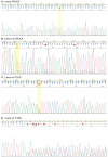
Sanger sequencing electropherograms of the OVAR79 cell line showing mutations in the PIK3CA and PTEN genes. (A) A single-nucleotide substitution in exon 1 (c.338T>C) leading to a codon change (CTC>CCC) and an amino acid substitution (Leu113Pro). (B) An indel mutation in exon 9 with a GT deletion and C insertion at position 1658–1659 leading to a codon change (AGT>ACC) and a frameshift. (C) A single-nucleotide variant in intron 2, rs1903858 at chr10:87893929. (D) An indel mutation around exon 4, rs1426397261, due to a duplication of ATACATATT to ATACATATTATACATATT, located at chr10:87931188-87931196.
Figure 3
Figure 3
Karyotype of the OVAR79 cell line. Regions of gain are indicated in green, regions of loss in red, and copy-neutral losses of heterozygosity (cnLOH) are shown in light blue.
Figure 4
Figure 4
(A) RT-qPCR analysis of mRNA expression levels of epithelial, mesenchymal, and stemness markers in OVAR79, SKOV3, and OVCAR3 cell lines. Bars represent the relative expression levels of each marker normalized to GAPDH (n = 3 biologically independent experiments). Data represent the mean values ± SEM. (B) Flow cytometry analysis of ovarian cancer markers in OVAR79, SKOV3, and OVCAR3 cell lines. The green color represents the antibody of interest, while the blue color corresponds to the respective isotype control for each antibody. Graphs display marker intensity on the x-axis and the percentage of the cell population on the y-axis (%). (C) Dose–response curves obtained by MTT assay of OVAR79, OVCAR3, and SKOV3 cells that were treated with different concentrations of cisplatin, carboplatin, or paclitaxel for 48 h. The data represent the mean values ± SD (n = 3 biologically independent replicates). IC50 values were determined by fitting a normalized dose–response model to the data using nonlinear regression in GraphPad Prism 8.0 software.

Similar articles

References

    1. Webb P.M., Jordan S.J. Epidemiology of Epithelial Ovarian Cancer. Best Pract. Res. Clin. Obstet. Gynaecol. 2017;41:3–14. - PubMed
    1. Kuroki L., Guntupalli S.R. Treatment of Epithelial Ovarian Cancer. BMJ. 2020;371:m3773. doi: 10.1136/bmj.m3773. - DOI - PubMed
    1. Ghose A., Bolina A., Mahajan I., Raza S.A., Clarke M., Pal A., Sanchez E., Rallis K.S., Boussios S. Hereditary Ovarian Cancer: Towards a Cost-Effective Prevention Strategy. Int. J. Environ. Res. Public Health. 2022;19:12057. doi: 10.3390/ijerph191912057. - DOI - PMC - PubMed
    1. Rojas V., Hirshfield K.M., Ganesan S., Rodriguez-Rodriguez L. Molecular Characterization of Epithelial Ovarian Cancer: Implications for Diagnosis and Treatment. Int. J. Mol. Sci. 2016;17:2113. doi: 10.3390/ijms17122113. - DOI - PMC - PubMed
    1. Cancer of the Ovary—Cancer Stat Facts. [(accessed on 25 September 2024)]; Available online: https://seer.cancer.gov/statfacts/html/ovary.html.

MeSH terms

Substances