. 2024 Dec 10;25(24):13251.
doi: 10.3390/ijms252413251.

DEAD/H Box 5 (DDX5) Augments E2F1-Induced Cell Death Independent of the Tumor Suppressor p53

Affiliations

DEAD/H Box 5 (DDX5) Augments E2F1-Induced Cell Death Independent of the Tumor Suppressor p53

Rinka Nakajima et al. Int J Mol Sci. .

Abstract

In almost all cancers, the p53 pathway is disabled and cancer cells survive. Hence, it is crucially important to induce cell death independent of p53 in the treatment of cancers. The transcription factor E2F1 is controlled by binding of the tumor suppressor pRB, and induces apoptosis by activating the ARF gene, an upstream activator of p53, when deregulated from pRB by loss of pRB function. Deregulated E2F1 can also induce apoptosis, independent of p53, via other targets such as TAp73 and BIM. We searched for novel E2F1-interacting proteins and identified the RNA helicase DEAD/H box 5 (DDX5), which also functions as a transcriptional coactivator. In contrast to the reported growth-promoting roles of DDX5, we show that DDX5 suppresses cell growth and survival by augmentation of deregulated E2F1 activity. Over-expression of DDX5 enhanced E2F1 induction of tumor suppressor gene expression and cell death. Conversely, shRNA-mediated knockdown of DDX5 compromised both. Moreover, DDX5 modulated E2F1-mediated cell death independent of p53, for which DDX5 also functions as a coactivator. Since p53 function is disabled in almost all cancers, these results underscore the roles of DDX5 in E2F1-mediated induction of cell death, independent of p53, and represent novel aspects for the treatment of p53-disabled cancer cells.

Keywords: ARF; BIM; DDX5; E2F1; TAp73; cell death; p53; pRB.


Conflict of interest statement

The authors declare no conflicts of interest.

Figures

Figure 1
Figure 1
DDX5 enhances over-expressed E2F1 activity in Hela cells. (A) Doxycyclin-inducible 3xFLAG-E2F1 HeLa cell lines were established by stably introducing T-REx-HeLa cells with pcDNA4/TO-3xFLAG-E2F1-puro. Puromycin-resistant clones were screened for transactivation ability by reporter assay using an ARF promoter-driven luciferase reporter. Fold activations by addition of Doxycyclin of three representative clones are shown. As a positive control, T-REx HeLa cells were transiently transfected with pcDNA4/TO-3xFLAG-E2F1-puro (T-REx + E2F1). (B) Expression of 3xFLAG-E2F1 in the three representative clones with or without Doxycyclin treatment was examined by western blot analysis using anti-E2F1 antibody. (C) Ten mg of nuclear extracts from T-REx-HeLa-3xFLAG-E2F1 with or without Doxycyclin treatment were immunoprecipitated with anti-FLAG antibody-conjugated agarose beads. Eluates were resolved by SDS-PAGE followed by silver staining. (D) DDX5 enhanced over-expressed E2F1 activity in HeLa cells. HeLa cells were transfected with ARF or TAp73 reporter plasmids and E2F1 expression plasmid along with increasing amounts (0, 20, 100, 500 ng) of DDX5 expression vector. Fold activations by E2F1 are shown. ***: p < 0.01.
Figure 2
Figure 2
DDX5 enhances E2F1 induction of endogenous target gene expression and cell death in normal cells. (A) DDX5 enhances over-expressed E2F1 activity in human normal fibroblasts (HFF). HFFs were transfected with the ARF or TAp73 reporter plasmid and E2F1 expression plasmid with increasing amounts (0, 125, 250, 500 ng) of DDX5 expression vector. Fold activations by E2F1 are shown. ***: p < 0.01. (B) DDX5 enhances endogenous deregulated E2F activity in normal cells. HFFs were transfected with the ARF reporter plasmid with E1a and DDX5 expression vectors. ***: p < 0.01. (C) Enhancement by DDX5 is through deregulated E2F1 activity. HFFs were transfected with a reporter plasmid possessing 3 tandem repeats of the ARF promoter E2F-responsive element (EREA (WT)), or its E2F binding site mutant (EREA (mt)), upstream of an SV40 core promoter (pGL3-Promoter), along with E2F1 and DDX5 expression vectors. pGL3-Promoter was used as a negative control. Fold induction by E2F1 is shown. ***: p < 0.01. (D) Enhancement of over-expressed E2F1 activity by DDX5 does not depend on helicase activity. Effects of helicase mutant DDX5(K144R) on E2F1 activation of the ARF (left panels) and TAp73 (right panels) promoters were examined at 500 ng of expression vector in as in (A). **: p < 0.05, ***: p < 0.01. (E) DDX5 enhances E2F1 induction of endogenous gene expression. HFFs were infected with Ad-FLAG-E2F1 (multiplicity of infection (MOI): 20) along with or without Ad-DDX5 or Ad-DDX5(K144R) (MOI: 50). The cells were further cultured for 48 h in the absence of serum and harvested. mRNA levels of ARF, BIM, and Aspp1 genes were examined by qRT-PCR. **: p < 0.05, ***: p < 0.01. (F) DDX5 enhances E2F1 induction of cell death. HFFs were infected with Ad-FLAG-E2F1 (MOI: 20) along with or without Ad-DDX5 or Ad-DDX5(K144R) (MOI: 50). The cells were cultured for 3 days and harvested. The percentage of dead cells was determined by FACS analysis as those with subG1 DNA content. ***: p < 0.01. (G) Expression of E2F1 and DDX5 protein levels were determined by western blot analysis under the same condition as in (E). β-actin was used as an internal control. (H) Expression levels of FLAG-E2F1 and DDX5 together with ARF, p53, and TAp73 were examined by western blot analysis under the same conditions as above. β-actin was used as an internal control. ***: p < 0.01. (I) DDX5 enhances endogenous deregulated E2F activity. HFFs were infected with Ad-12SE1a(∆2–11) (MOI: 200) along with or without Ad-DDX5 (MOI: 50). The cells were further cultured for 48 h in the absence of serum and harvested. mRNA levels of ARF, BIM, and Aspp1 genes were examined by qRT-PCR. **: p < 0.05, ***: p < 0.01. (J) Expression levels of E1a and DDX5 together with E2F1 were examined by western blot analysis.
Figure 3
Figure 3
Knockdown of DDX5 expression reduces E2F1-induction of tumor suppressor gene expression and cell death in normal cells. (A) Knockdown effects of shRNA against DDX5 (shDDX5) in HFFs. HFFs were infected with Ad-shDDX5-1, Ad-shDDX5-2, or control virus (MOI: 20), and cultured for 72 h. The cells were then infected with Ad-FLAG-E2F1 or control virus (MOI: 20), further cultured for 24 h, and harvested. Expression of DDX5 and FLAG-E2F1 protein levels were determined by western blotting. β-actin was used as an internal control. **: p < 0.05, ***: p < 0.01. (B) Knockdown of DDX5 reduced E2F1 induction of ARF, BIM, and Aspp1 gene expression in HFFs. The cells were infected with Ad-shDDX5-1 or -2 and FLAG-E2F1 in the same way as above. mRNA levels of the ARF, BIM, and Aspp1 genes were examined by qRT-PCR. Knockdown of DDX5 was confirmed at mRNA levels by qRT-PCR. **: p < 0.05, ***: p < 0.01. (C) Knockdown of DDX5 decreased E2F1 induction of cell death in HFFs. HFFs were infected with shDDX5-1 or -2 and FLAG-E2F1 in the same way as above, cultured for 3 days and harvested. The percentage of dead cells was determined by FACS analysis as those with subG1 DNA content. ***: p < 0.01.
Figure 4
Figure 4
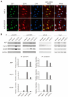
Deregulated E2F1 recruits DDX5 to target genes. (A) DDX5 and E2F1 are co-localized in the nucleus. HFFs were infected with Ad-12SE1a(∆2–11) (upper panel) or Ad-FLAG-E2F1 (lower panel), and further cultured for 48 h in the absence of serum. Localization of DDX5 and E2F1 was examined by confocal laser microscopy. Yellow color in (E2F1+DDX5 merge) indicates that both proteins fluorophores are in close proximity, suggesting colocalization. (B) DDX5 was recruited to E2F1 target genes in HFFs. HFFs were starved of serum, restimulated with serum, or deregulated E2F1 activity was generated by over-expression of E2F1 or expression of E1a by adenovirus-mediated gene transfer. ChIP assay was performed using E2F1 and DDX5-specific antibodies. Anti-hemagglutinin (HA) antibody and the glyceraldehyde 3-phosphate dehydrogenase (GAPDH) gene served as negative controls. Intensities of bands were measured by ImageJ and presented as relative intensities.
Figure 5
Figure 5
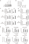
DDX5 enhances E2F1 induction of tumor suppressor gene expression and cell death in normal cells transduced with dominant negative mutants of p53. (A) HFFs were stably transduced with DNp53s. HFFs were immortalized by stably introducing hTERT and subsequently infected with either of two DNp53s, R175H or R273H, using retroviral vectors. Expression of endogenous p53 protein levels in the cells was examined by western blot analysis. β-actin was used as an internal control. (B) Dominant negative effects of DNp53s. The cells were infected with Ad-p53 (MOI: 50), cultured for 24 h and harvested. The levels of p53AIP1 gene expression were examined by qRT-PCR. ***: p < 0.01. hTEPT immortalized HFFs were used as control cells. (C) DDX5 enhanced E2F1 induction of tumor suppressor genes in DNp53-introduced HFFs. The cells were infected with Ad-FLAG-E2F1 and Ad-DDX5, cultured for 48 h and harvested. The levels of BIM and Aspp1 genes expression were examined by qRT-PCR. **: p < 0.05, ***: p < 0.01. (D) DDX5 enhanced E2F1 induction of cell death in DNp53-transduced HFFs. The cells were infected with Ad-FLAG-E2F1 and Ad-DDX5, cultured for 72 h and harvested. Percentage of dead cells was examined by FACS analysis as those with subG1 DNA content. ***: p < 0.01. (E) Knockdown of BIM by shRNA decreased E2F1-induced cell death in DNp53-introduced HFFs. The cells were infected with Ad-shBIM-1 or -2 (MOI: 2) and Ad-FLAG-E2F1 (MOI: 50), cultured for 3 days and harvested. The percentage of dead cells was determined by FACS analysis as those with subG1 DNA content. ***: p < 0.01. (F) Knockdown effects of shBIMs examined by qRT-PCR under the same condition as above. **: p < 0.05, ***: p < 0.01.
Figure 6
Figure 6
DDX5 enhances E2F1-mediated p53-independent cell death in cancer cells. (A,B) DDX5 enhances E2F1 induction of tumor suppressor genes expression in p53 null cancer cells. H1299 (A) and Saos-2 (B) cells were infected with Ad-FLAG-E2F1 (MOI: 2) along with or without Ad-DDX5 (MOI: 5). The cells were further cultured for 24 h and harvested. mRNA levels of BIM and Aspp1 genes were examined by qRT-PCT. **: p < 0.05, ***: p < 0.01. (C) DDX5 enhances E2F1-induced cell death in Saos-2 cells. Saos-2 cells were infected in the same way as above, further cultured for 2 days and harvested. Percentage of dead cells was determined by FACS analysis as those with subG1 DNA content. ***: p < 0.01. (D) Knockdown of DDX5 decreases E2F1 induction of tumor suppressor gene expression in Saos-2 cells. Cells were infected with Ad-shDDX5-1 (MOI: 1) or -2 (MOI: 2), and cultured for 72 h. The cells were then infected with Ad-FLAG-E2F1 (MOI: 2) further cultured for 24 h and harvested. The levels of BIM and Aspp1 gene expression, and knockdown of DDX5 gene expression were examined by qRT-PCR. **: p < 0.05, ***: p < 0.01. (E) Knockdown of DDX5 decreases E2F1-induced cell death in Saos-2 cells. The cells were infected in the same way as above, cultured for 3 days and harvested. The percentage of dead cells was examined as above. ***: p < 0.01. (F,G) Over-expression of E2F1 induces cell death in p53-disabled cancer cells. H1299 (F) and Saos-2 (G) cells were infected with Ad-shBIM-1 (MOI: 1) or -2 (MOI: 2) and Ad-FLAG-E2F1 (MOI: 2), further cultured for 3 days and harvested. Percentage of dead cells was examined as above (left panels). Knockdown of BIM by shRNA was confirmed by qRT-PCR (right panels). **: p < 0.05, ***: p < 0.01.
Figure 7
Figure 7
A model for the role of DDX5 in E2F1-mediated tumor suppressor gene expression and cell death. DDX5 enhances E2F1 induction of tumor suppressor gene expression, thereby enhancing E2F1-mediated cell death, independent of p53.

Similar articles

Cited by

References

    1. Trimarchi J.M., Lees J.A. Sibling rivalry in the E2F family. Nat. Rev. Mol. Cell Biol. 2002;3:11–20. doi: 10.1038/nrm714. - DOI - PubMed
    1. Iaquinta P.J., Lees J.A. Life and death decisions by the E2F transcription factors. Curr. Opin. Cell Biol. 2007;19:649–657. doi: 10.1016/j.ceb.2007.10.006. - DOI - PMC - PubMed
    1. Chen H.Z., Tsai S.Y., Leone G. Emerging roles of E2Fs in cancer: An exit from cell cycle control. Nat. Rev. Cancer. 2009;9:785–797. doi: 10.1038/nrc2696. - DOI - PMC - PubMed
    1. Polager S., Ginsberg D. p53 and E2f: Partners in life and death. Nat. Rev. Cancer. 2009;9:738–748. doi: 10.1038/nrc2718. - DOI - PubMed
    1. Poppy Roworth A., Ghari F., La Thangue N.B. To live or let die—Complexity within the E2F1 pathway. Mol. Cell. Oncol. 2015;2:e970480. doi: 10.4161/23723548.2014.970480. - DOI - PMC - PubMed

MeSH terms

LinkOut - more resources