Review
. 2024 Dec 11;25(24):13279.
doi: 10.3390/ijms252413279.

Metal-Dependent Cell Death in Renal Fibrosis: Now and in the Future

Affiliations
Review

Metal-Dependent Cell Death in Renal Fibrosis: Now and in the Future

Te Li et al. Int J Mol Sci. .

Abstract

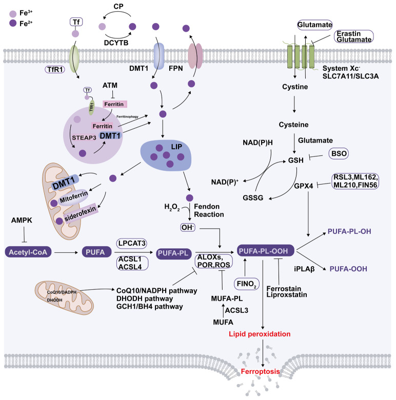
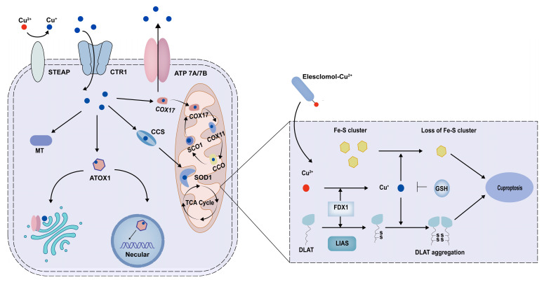
Renal fibrosis is a common final pathway underlying nearly almost all progressive kidney diseases. Metal ions are essential trace elements in organisms and are involved in important physiological activities. However, aberrations in intracellular metal ion metabolism may disrupt homeostasis, causing cell death and increasing susceptibility to various diseases. Accumulating evidence suggests a complex association between metal-dependent cell death and renal fibrosis. In this article, we provide a comprehensive overview of the specific molecular mechanisms of metal-dependent cell death and their crosstalk, up-to-date evidence supporting their role in renal fibrosis, therapeutic targeting strategies, and research needs, aiming to offer a rationale for future clinical treatment of renal fibrosis.

Keywords: cell death; cuproptosis; ferroptosis; metal ions; renal fibrosis; zinc.


Conflict of interest statement

The authors declare no conflicts of interest.

Figures

Figure 1
Figure 1
Iron metabolism and ferroptosis. Extracellular irons predominantly exist in the form of Fe3+. Fe3+ is first reduced to Fe2+ by ferrireductases, transporting it into the cells via DMT1. Intracellular Fe2+ is exported via ferroportin located on the basolateral membrane and oxidized to Fe3+ with the help of ceruloplasmin, allowing it to bind to transferrin for circulation. The majority of cellular iron is stored in ferritin, while a portion is in LIPs. Mitoferrin, siderofexin, and DMT1 can transport Fe2+ stored from the LIPs to the mitochondria. Excess intracellular iron from the LIP can be stored in lysosomes as ferritin. Iron is typically present as Fe2+ within lysosomes due to their acidic and reducing conditions. When intracellular iron ions are reduced, the autophagic degradation of ferritin in lysosomes can release divalent iron to replenish LIP. Ferroptosis is characterized by the depletion of intracellular glutathione and decreased activity of GPX4, which leads to the accumulation of unmetabolized lipid peroxides and increased ROS production. Membrane damage is also a result of lipid peroxidation. Moreover, the FSP1/CoQ10/NAD(P)H axis, the GCH1/BH4 axis, and the DHODH pathways suppress ferroptosis in parallel with the GPX4-dependent pathway. ALOXs, lipoxygenases; AMPK, adenosine-monophosphate-activated protein kinase; ACSL, acyl-CoA synthetase long-chain family member; ATM, ATM serine/threonine kinase; BSO, buthionine sulfoximine; CP, ceruloplasmin; CoA, coenzyme A; CoQ10, coenzyme CQ10; DCYTB, duodenal cytochrome B; DHODH, dihydroorotate dehydrogenase (quinone); DMT1, divalent metal-ion transporter 1; FPN, ferroportin; FSP1, ferroptosis suppressor protein 1; GCH1, GTP cyclohy drolase 1; GCLC, glutamate-cysteine ligase catalytic subunit; Glu, glutamate; GPX4, glutathione peroxidase 4; GSH, glutathione; iPLA2b, phospholipase A2 group VI; LIP, labile iron pools; LPCAT3, lysophosphatidylcholine acyltransferase 3; MUFA, monounsaturated fatty acid; NADPH, reduced nicotinamide adenine dinucleotide phosphate; PL, phospholipid; POR, Cytochrome p450 oxidoreductase; PUFA, polyunsaturated fatty acid; PUFA-PL-OOH, phospholipid with peroxidized polyunsaturated fatty acyl tail; ROS, reactive oxygen species; System xc-, sodium-independent, anionic amino acid transport system; ATSTEAP, six-transmembrane epithelial antigen of the prostat; Tf, transferrin; TfR1, transferrin receptor protein 1.
Figure 2
Figure 2
Copper metabolism and cuproptosis. Extracellular copper ions predominantly exist in the form of Cu2+. Cu2+ is reduced to Cu+ via the action of STEAP on the intestinal cell membrane. The cellular uptake of Cu+ is facilitated by CTR1, whereas its export is mediated by ATP7A and ATP7B. Intracellularly, Cu+ is directed to various subcellular organelles for bioavailability by copper-binding proteins, including COX17, CCS, and ATOX1. Additionally, the binding of MT to copper can mitigate the cytotoxicity associated with copper excess. Elesclomol binds Cu2+ in the extracellular environment and transports it to mitochondria. Cu+ directly binds to DLAT, promoting protein acylation and inducing the degradation of Fe-S cluster proteins, which ultimately induces proteotoxic stress and cell death. GSH serves as a thiol-containing copper chelator that blocks cuproptosis. STEAP, six-transmembrane epithelial antigen of the prostate; CTR1, copper affinity transporter 1; ATP7A/7B, ATPases of the 7A and 7B; COX17, cytochrome C oxidase 17; SCO1, synthesis cytochrome c oxidase 1; CCO, cytochrome c oxidase; SOD1, superoxide dismutase 1; CCS, copper chaperone for superoxide; ATOX1, antioxidant 1 copper chaperone; MT, metal-ion transporter; TCA cycle, mitochondrial tricarboxylic acid cycle; FDX1, ferredoxin 1; DLAT, dihydrolipoamide S-acetyltransferase; LIAS, lipoic acid synthetase; GSH, glutathione. Fe-S, iron-sulfur.

Similar articles

  • Endoplasmic Reticulum Stress-Mediated Cell Death in Renal Fibrosis.
    Guo S, Tong Y, Li T, Yang K, Gao W, Peng F, Zou X. Guo S, et al. Biomolecules. 2024 Jul 28;14(8):919. doi: 10.3390/biom14080919. Biomolecules. 2024. PMID: 39199307 Free PMC article. Review.
  • Adiponectin in renal fibrosis.
    Jing H, Tang S, Lin S, Liao M, Chen H, Fan Y, Zhou J. Jing H, et al. Aging (Albany NY). 2020 Feb 17;12(5):4660-4672. doi: 10.18632/aging.102811. Epub 2020 Feb 17. Aging (Albany NY). 2020. PMID: 32065783 Free PMC article. Review.
  • Iron and ferroptosis in kidney disease: molecular and metabolic mechanisms.
    Wang W, Chen J, Zhan L, Zou H, Wang L, Guo M, Gao H, Xu J, Wu W. Wang W, et al. Front Immunol. 2025 Feb 5;16:1531577. doi: 10.3389/fimmu.2025.1531577. eCollection 2025. Front Immunol. 2025. PMID: 39975561 Free PMC article. Review.
  • Kidney Fibrosis and Matrix Metalloproteinases (MMPs).
    La Russa A, Serra R, Faga T, Crugliano G, Bonelli A, Coppolino G, Bolignano D, Battaglia Y, Ielapi N, Costa D, Michael A, Andreucci M. La Russa A, et al. Front Biosci (Landmark Ed). 2024 May 16;29(5):192. doi: 10.31083/j.fbl2905192. Front Biosci (Landmark Ed). 2024. PMID: 38812325 Review.
  • Ferroptosis in renal fibrosis: a mini-review.
    Yang SQ, Zhao X, Zhang J, Liao DY, Wang YH, Wang YG. Yang SQ, et al. J Drug Target. 2024 Aug;32(7):785-793. doi: 10.1080/1061186X.2024.2353363. Epub 2024 May 14. J Drug Target. 2024. PMID: 38721679 Review.

References

    1. Liu Y. Cellular and molecular mechanisms of renal fibrosis. Nat. Rev. Nephrol. 2011;7:684–696. doi: 10.1038/nrneph.2011.149. - DOI - PMC - PubMed
    1. Huang R., Fu P., Ma L. Kidney fibrosis: From mechanisms to therapeutic medicines. Signal Transduct. Target. Ther. 2023;8:129. doi: 10.1038/s41392-023-01379-7. - DOI - PMC - PubMed
    1. Humphreys B.D. Mechanisms of Renal Fibrosis. Annu. Rev. Physiol. 2018;80:309–326. doi: 10.1146/annurev-physiol-022516-034227. - DOI - PubMed
    1. Huang J., Tong Z., Xu T., Zhang M., Ma Y., Wang J., Zhou H., Sun L. Copper homeostasis in chronic kidney disease and its crosstalk with ferroptosis. Pharmacol. Res. 2024;202:107139. doi: 10.1016/j.phrs.2024.107139. - DOI - PubMed
    1. Jomova K., Makova M., Alomar S.Y., Alwasel S.H., Nepovimova E., Kuca K., Rhodes C.J., Valko M. Essential metals in health and disease. Chem.-Biol. Interact. 2022;367:110173. doi: 10.1016/j.cbi.2022.110173. - DOI - PubMed

LinkOut - more resources