Cellular and Molecular Effects of the Bruck Syndrome-Associated Mutation in the PLOD2 Gene
- PMID: 39769143
- PMCID: PMC11676324
- DOI: 10.3390/ijms252413379
Cellular and Molecular Effects of the Bruck Syndrome-Associated Mutation in the PLOD2 Gene
Abstract
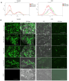
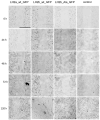
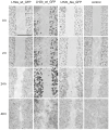
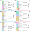
Bruck syndrome is a rare autosomal recessive disorder characterized by increased bone fragility and joint contractures similar to those in arthrogryposis and is known to be associated with mutations in the FKBP10 (FKBP prolyl isomerase 10) and PLOD2 (Procollagen-Lysine,2-Oxoglutarate 5-Dioxygenase 2) genes. These genes encode endoplasmic reticulum proteins that play an important role in the biosynthesis of type I collagen, which in turn affects the structure and strength of connective tissues and bones in the body. Mutations are associated with disturbances in both the primary collagen chain and its post-translational formation, but the mechanism by which mutations lead to Bruck syndrome phenotypes has not been determined, not only because of the small number of patients who come to the attention of researchers but also because of the lack of disease models. In our work, we investigated the cellular effects of two forms of the wild-type PLOD2 gene, as well as the PLOD2 gene with homozygous mutation c.1885A>G (p.Thr629Ala). The synthesized genetic constructs were transfected into HEK293 cell line and human skin fibroblasts (DF2 line). The localization of PLOD2 protein in cells and the effects caused by the expression of different isoforms-long, short, and long with mutation-were analyzed. In addition, the results of the transcriptome analysis of a patient with Bruck syndrome, in whom this mutation was detected, are presented.
Keywords: Bruck syndrome; PLOD2; cells; mutations; transfection.
Conflict of interest statement
The authors declare no conflicts of interest.
Figures


Similar articles
-
Loss of the long form of Plod2 phenocopies contractures of Bruck syndrome-osteogenesis imperfecta.J Bone Miner Res. 2024 Sep 2;39(9):1240-1252. doi: 10.1093/jbmr/zjae124. J Bone Miner Res. 2024. PMID: 39088537 Free PMC article.
-
Bruck syndrome 2 variant lacking congenital contractures and involving a novel compound heterozygous PLOD2 mutation.Bone. 2020 Jan;130:115047. doi: 10.1016/j.bone.2019.115047. Epub 2019 Aug 28. Bone. 2020. PMID: 31472299 Free PMC article.
-
Novel Mutations in PLOD2 Cause Rare Bruck Syndrome.Calcif Tissue Int. 2018 Mar;102(3):296-309. doi: 10.1007/s00223-017-0360-6. Epub 2017 Nov 24. Calcif Tissue Int. 2018. PMID: 29177700
-
Bruck syndrome - a rare syndrome of bone fragility and joint contracture and novel homozygous FKBP10 mutation.Endokrynol Pol. 2015;66(2):170-4. doi: 10.5603/EP.2015.0024. Endokrynol Pol. 2015. PMID: 25931047 Review.
-
Bone collagen: new clues to its mineralization mechanism from recessive osteogenesis imperfecta.Calcif Tissue Int. 2013 Oct;93(4):338-47. doi: 10.1007/s00223-013-9723-9. Epub 2013 Mar 19. Calcif Tissue Int. 2013. PMID: 23508630 Free PMC article. Review.
References
-
- Mumm S., Gottesman G.S., Wenkert D., Campeau P.M., Nenninger A., Huskey M., Bijanki V.N., Veis D.J., Barnes A.M., Marini J.C., et al. Bruck syndrome 2 variant lacking congenital contractures and involving a novel compound heterozygous PLOD2 mutation. Bone. 2020;130:115047. doi: 10.1016/j.bone.2019.115047. - DOI - PMC - PubMed
-
- Puig-Hervás M.T., Temtamy S., Aglan M., Valencia M., Martínez-Glez V., Ballesta-Martínez M.J., López-González V., Ashour A.M., Amr K., Pulido V., et al. Mutations in PLOD2 cause autosomal-recessive connective tissue disorders within the Bruck syndrome--osteogenesis imperfecta phenotypic spectrum. Hum. Mutat. 2012;33:1444–1449. doi: 10.1002/humu.22133. - DOI - PubMed
-
- Gistelinck C., Witten P.E., Huysseune A., Symoens S., Malfait F., Larionova D., Simoens P., Dierick M., Van Hoorebeke L., De Paepe A., et al. Loss of Type I Collagen Telopeptide Lysyl Hydroxylation Causes Musculoskeletal Abnormalities in a Zebrafish Model of Bruck Syndrome. J. Bone Miner. Res. 2016;31:1930–1942. doi: 10.1002/jbmr.2977. - DOI - PMC - PubMed
MeSH terms
Substances
Supplementary concepts
Grants and funding
LinkOut - more resources
Full Text Sources
Miscellaneous

