. 2024 Dec 13;25(24):13389.
doi: 10.3390/ijms252413389.

Inhibiting De Novo Biosynthesis of Ceramide by L-Cycloserine Can Prevent Light-Induced Retinal Degeneration in Albino BALB/c Mice

Affiliations

Inhibiting De Novo Biosynthesis of Ceramide by L-Cycloserine Can Prevent Light-Induced Retinal Degeneration in Albino BALB/c Mice

Faiza Tahia et al. Int J Mol Sci. .

Abstract

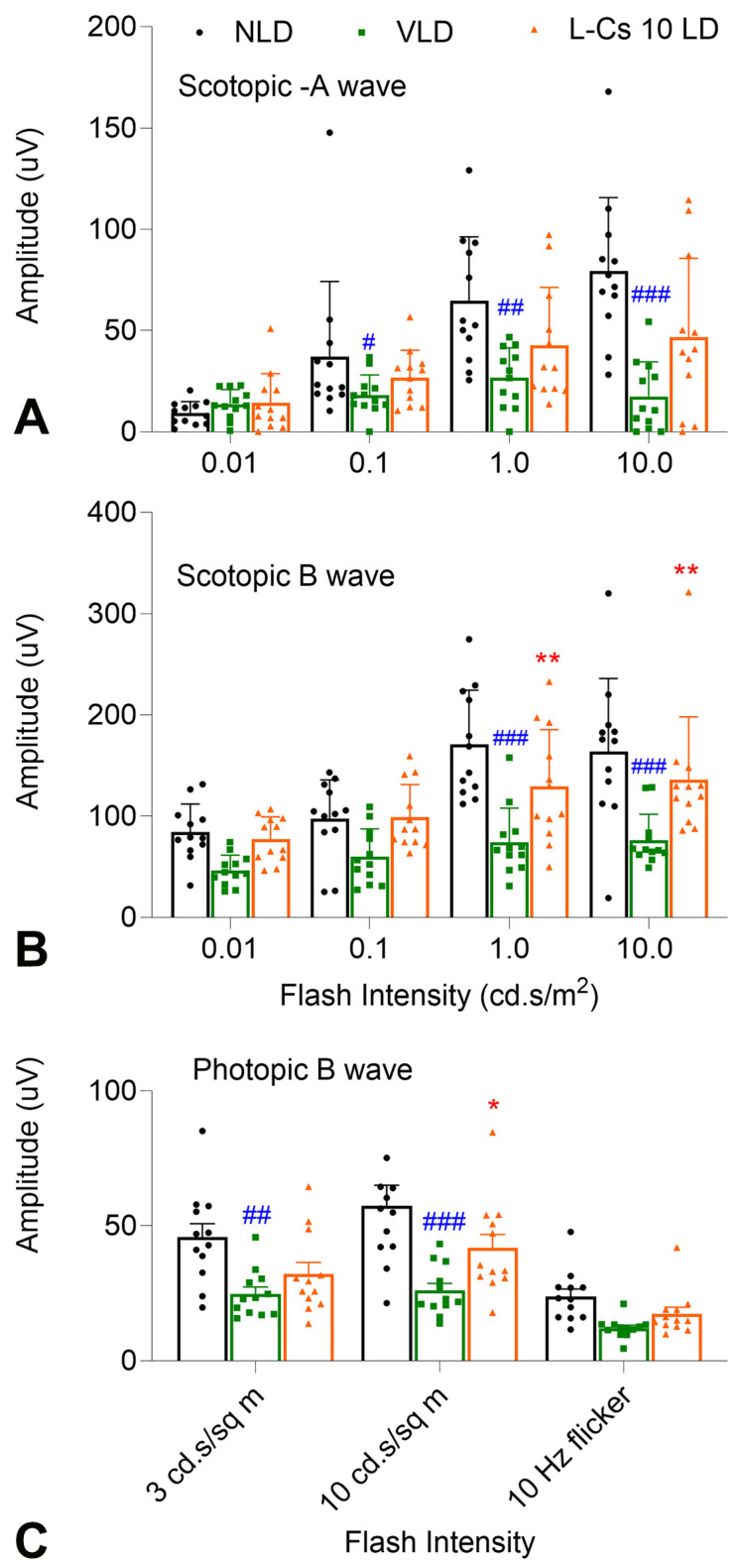
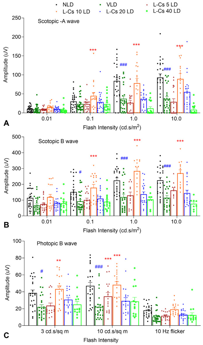
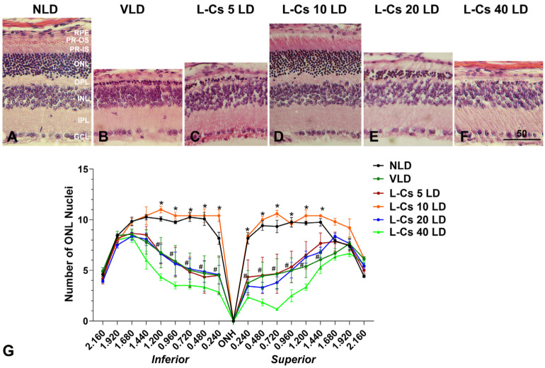
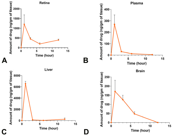
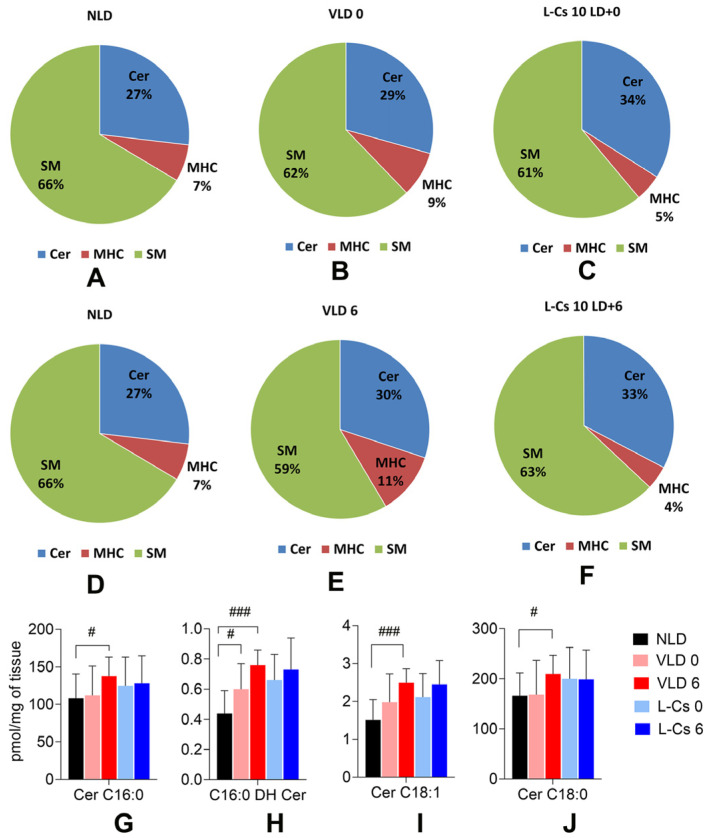
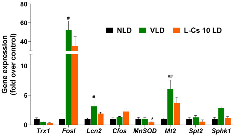
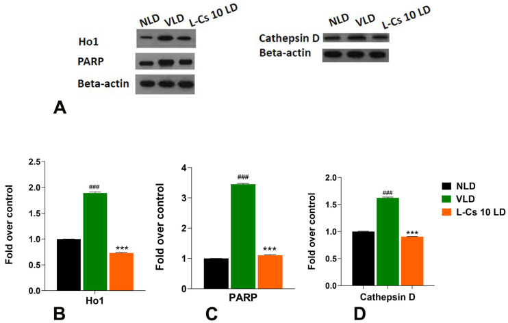
Retinal degenerative diseases lead to irreversible vision loss due to photoreceptor cell death, driven by complex genetic and environmental factors. Ceramide, a sphingolipid metabolite, emerges as a critical mediator in the apoptotic cascade associated with retinal degeneration. Our previous work demonstrated L-Cycloserine's ability to protect photoreceptor-derived cells from oxidative stress by inhibiting the de novo ceramide pathway and thus prompting further investigation on its effect in the in vivo retina. This study investigates the potential of L-Cycloserine to protect albino BALB/c mice against light-induced retinal degeneration (LIRD). L-Cycloserine, in an optimal dose, administered systemically 30 min before LIRD, was found to prevent photoreceptor cell death significantly from light-induced degeneration. We further determined the retinal bioavailability and pharmacokinetic behavior of L-Cycloserine, its effect on sphingolipid profile, expression of sphingolipid biosynthetic, and cell death-promoting genes and proteins from the retina to understand the underlying mechanisms. This study lays the groundwork for further preclinical and clinical investigations into L-Cycloserine's potential as a novel therapeutic in treating retinal degenerative diseases.

Keywords: BALB/c mice; L-Cycloserine; ceramide; light-induced retinal degeneration (LIRD); pharmacokinetics; photoreceptor cell death; retinal degeneration.


Conflict of interest statement

The authors declare no conflicts of interest.

Figures

Figure 1
Figure 1
L-Cycloserine protects mouse retina from LIRD. ERG analysis shows Scotopic a-wave (A), b-wave (B), and Photopic b-wave (C) from NLD: no light-damaged control group; VLD: vehicle-injected light-damaged group; L-Cs 10 LD: L-Cycloserine-treated (10 mg/kg) light-damaged group. (n = 6/group; values are mean ± SEM; * represents significance between VLD and L-Cs 10 LD; * p < 0.05, ** p < 0.01, by the student t test; # represents significance between NLD and VLD; # p < 0.05, ## p < 0.01, ### p < 0.001, by the student t test).
Figure 2
Figure 2
A 10 mg/kg dose of L-Cycloserine provides maximum protection of mouse retina from LIRD. ERG analysis shows Scotopic a-wave (A), b-wave (B), and Photopic b-wave (C) from NLD: no light-damaged control group; VLD: vehicle-injected light-damaged group; L-Cs 5 LD,10 LD, 20 LD, and 40 LD: L-Cycloserine-treated (5, 10, 20, and 40 mg/kg, respectively) light-damaged group. n = 13 (NLD, VLD); n = 9 (L-Cs 5 LD, L-Cs 10 LD); n = 10 (L-Cs 20 LD); n = 8 (L-Cs 40 LD); values are mean ± SEM; * represents significance between VLD and L-Cycloserine treated group; ** p < 0.01, *** p < 0.001, by the student t test; # represents significance between NLD and VLD; # p < 0.05, ### p < 0.001, by the student t test).
Figure 3
Figure 3
A 10 mg/kg dose of L-Cycloserine provides maximum protection of mouse retina from LIRD. Representative retinal histological sections from each treatment: (A): NLD, no light-damaged control; (B): VLD, vehicle-injected light-damaged group; (CF): L-Cs 5 LD,10 LD, 20 LD, and 40 LD, represents L-Cycloserine-treated (5, 10, 20, and 40 mg/kg, respectively) light-damaged group. Scale bar in F represents 50 microns. (G) Quantitative morphometric measurement of ONL nuclei count from H and E-stained slides. n = 12 (NLD); n = 16 (VLD); n = 10 (L-Cs 5 LD); n = 13 (L-Cs 10 LD); n = 14 (L-Cs 20 LD); n = 11 (L-Cs 40 LD). Values are mean ± SEM; * represents significance between VLD and L-Cycloserine treated group; * p < 0.001, by the student t test; # represents significance between NLD and VLD; # p < 0.001, by the student t test. Abbreviations for retinal section: RPE, retinal pigment epithelium; PR, photoreceptors; OS, outer segments; IS, inner segments; ONL, outer nuclear layer; OPL, outer plexiform layer; INL, inner nuclear layer; IPL, inner plexiform layer; GCL, ganglion cell layer.
Figure 4
Figure 4
Pharmacokinetic profile of L-Cycloserine in plasma and various tissues. L-cycloserine was intraperitoneally injected at a dose of 20 mg/kg and quantified using LC-MS/MS in retina (A), plasma (B), liver (C), and brain (D) at various time points after administration. The results are expressed as a mean ± SEM (n = 7 for each time point).
Figure 5
Figure 5
Light damage and L-Cycloserine treatment alter ceramide and other sphingolipid levels in light-damaged BALB/c mice retina. (AF): Analysis of major sphingolipid levels by absolute value (pmol/mg of tissue) showing the total composition of ceramide (Cer), sphingomyelin (SM), and monohexosylceramide (MHC) percent in BALB/c mice retina (n = 10/group). Analysis of selected ceramide species by absolute value (pmol/mg of tissue) in BALB/c mice retina at 0 h and 6 h after light damage (GJ). (n = 10/group, values are mean ± SEM; # represents significance between NLD and VLD; # p < 0.05, ### p < 0.001, by student t-test). NLD: no light-damaged control group, VLD 0: vehicle-injected light-damaged group at 0 h after light damage, L-Cs 0: L-Cycloserine (10 mg/kg) treated light-damaged group at 0 h after light damage, VLD 6: vehicle-injected light-damaged group at 6 h after light damage and L-Cs 6: L-Cycloserine (10 mg/kg) treated light-damaged group at 6 h after light damage.
Figure 6
Figure 6
L-Cycloserine modulates retinal gene expression in light-damaged BALB/c mice. Quantitative analysis of gene expression of no light-damaged control (NLD), vehicle-injected light-damaged (VLD), and L-Cycloserine (10 mg/kg) treated light-damaged (L-Cs 10 LD) retina 0 h after light damage. Expression values (±SEM) are presented against fold change over control value (NLD), which was set to 1.0 (n = 6; # represents significance between NLD and VLD; # p < 0.05, ## p < 0.01, by the student t test; * represents significance between VLD and L-Cs 10 LD; * p < 0.05, by the student t test).
Figure 7
Figure 7
L-Cycloserine modulates expression of retinal proteins in light-damaged BALB/c mice. (A): Expression and quantification of heme oxygenase 1 (Ho1), Poly (ADP-ribose) polymerase (PARP) and Cathepsin D in BALB/c retina from no light-damaged control (NLD), vehicle-injected light-damaged (VLD) and L-Cycloserine (10 mg/kg) treated light-damaged (L-Cs 10 LD) group, 0 h after light damage. Quantification of (B): Ho1 (C) PARP (D): Cathepsin D in retinal tissue obtained with densitometric analysis and normalized with β-actin. (n = 3; # represents significance between NLD and VLD; ### p < 0.001, by student t-test; * represents significance between VLD and L-Cs 10 LD; *** p < 0.001, by student t-test).

Similar articles

Cited by

References

    1. Bhattacharyya A. The detrimental effects of progression of retinal degeneration in the visual cortex. Front. Cell. Neurosci. 2022;16:904175. doi: 10.3389/fncel.2022.904175. - DOI - PMC - PubMed
    1. Wert K.J., Lin J.H., Tsang S.H. General pathophysiology in retinal degeneration. Dev. Ophthalmol. 2014;53:33–43. doi: 10.1159/000357294. - DOI - PMC - PubMed
    1. Daiger S.P. Retinal Information Network. 1996–2022. [(accessed on 1 March 2024)]. Available online: https://web.sph.uth.edu/RetNet/
    1. John M.C., Quinn J., Hu M.L., Cehajic-Kapetanovic J., Xue K. Gene-agnostic therapeutic approaches for inherited retinal degenerations. Front. Mol. Neurosci. 2022;15:1068185. doi: 10.3389/fnmol.2022.1068185. - DOI - PMC - PubMed
    1. Ohanian J., Ohanian V. Sphingolipids in mammalian cell signalling. Cell. Mol. Life Sci. CMLS. 2001;58:2053–2068. doi: 10.1007/PL00000836. - DOI - PMC - PubMed

LinkOut - more resources