. 2024 Dec 14;25(24):13406.
doi: 10.3390/ijms252413406.

Exploring the Osteoinductive Potential of Bacterial Pyomelanin Derived from Pseudomonas aeruginosa in a Human Osteoblast Model

Affiliations

Exploring the Osteoinductive Potential of Bacterial Pyomelanin Derived from Pseudomonas aeruginosa in a Human Osteoblast Model

Mateusz M Urbaniak et al. Int J Mol Sci. .

Abstract

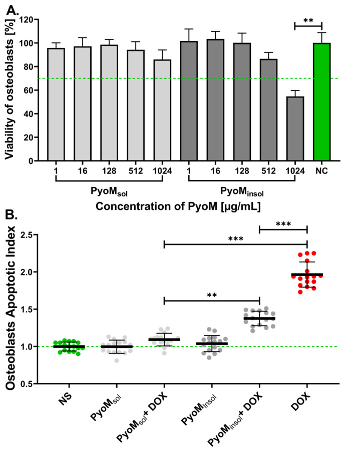
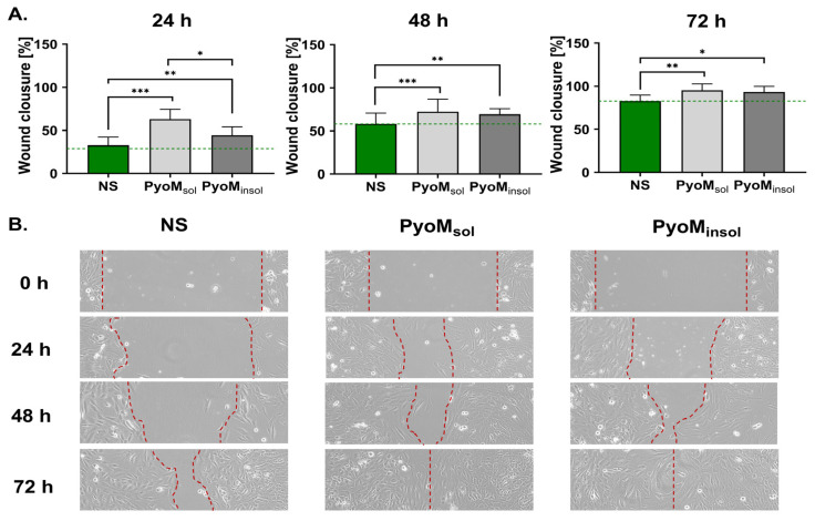
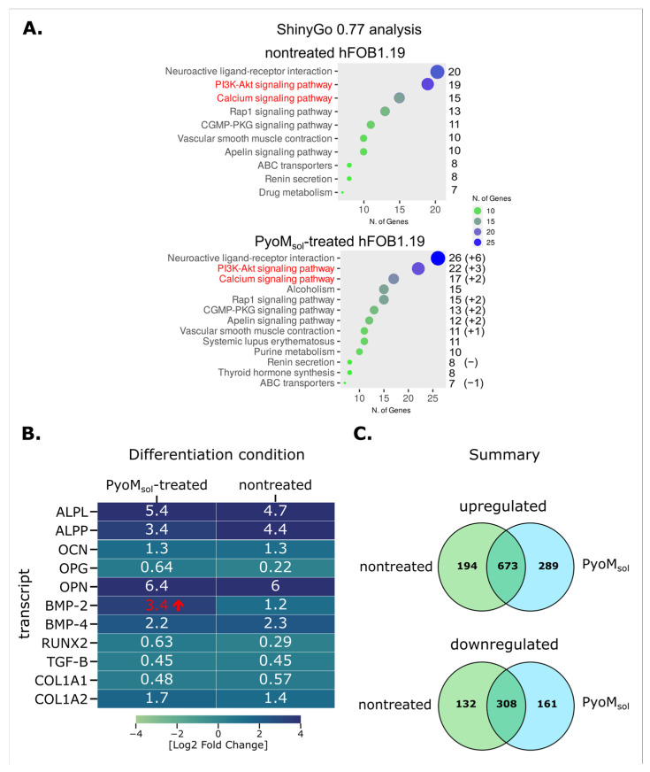
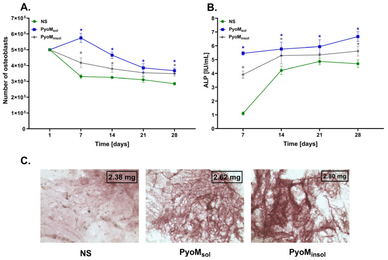
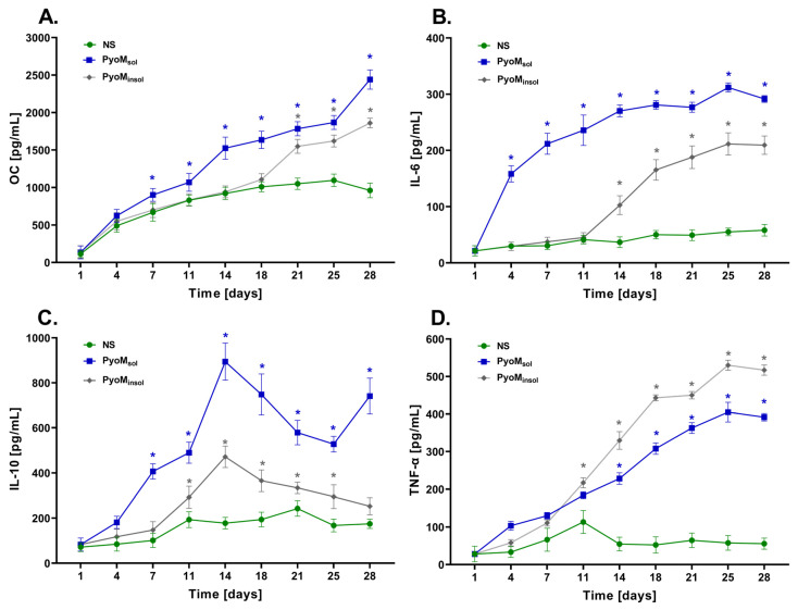
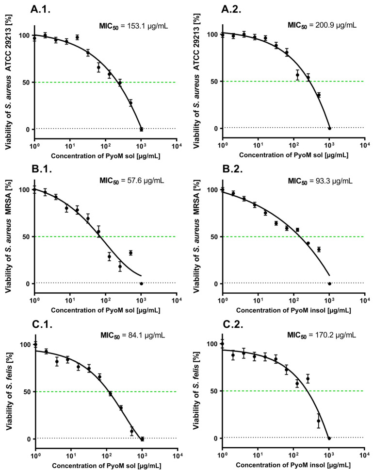
Alkaptonuria (AKU) is a genetically determined disease associated with disorders of tyrosine metabolism. In AKU, the deposition of homogentisic acid polymers contributes to the pathological ossification of cartilage tissue. The controlled use of biomimetics similar to deposits observed in cartilage during AKU potentially may serve the development of new bone regeneration therapy based on the activation of osteoblasts. The proposed biomimetic is pyomelanin (PyoM), a polymeric biomacromolecule synthesized by Pseudomonas aeruginosa. This work presents comprehensive data on the osteoinductive, pro-regenerative, and antibacterial properties, as well as the cytocompatibility, of water-soluble (PyoMsol) or water-insoluble (PyoMinsol) PyoM. Both variants of PyoM support osteoinductive processes as well as the maturation of osteoblasts in cell cultures in vitro due to the upregulation of bone-formation markers, osteocalcin (OC), and alkaline phosphatase (ALP). Furthermore, the cytokines involved in these processes were elevated in cell cultures of osteoblasts exposed to PyoM: tumor necrosis factor (TNF)-α, interleukin (IL)-6, and IL-10. The PyoM variants are cytocompatible in a wide concentration range and limit the doxorubicin-induced apoptosis of osteoblasts. This cytoprotective PyoM activity is correlated with an increased migration of osteoblasts. Moreover, PyoMsol and PyoMinsol exhibit antibacterial activity against staphylococci isolated from infected bones. The osteoinductive, pro-regenerative, and antiapoptotic effects achieved through PyoM stimulation prompt the development of new biocomposites modified with this bacterial biopolymer for medical use.

Keywords: bone regeneration; osteoblast; osteoinduction; pyomelanin.


Conflict of interest statement

The authors declare no conflicts of interest. The funders had no role in the design of the study; in the collection, analyses, or interpretation of data; in the writing of the manuscript; or in the decision to publish the results.

Figures

Figure 1
Figure 1
(A) The percentage of viable hFOB 1.19 osteoblasts after 24 h exposure to different concentrations of the water-soluble (PyoMsol) or water-insoluble pyomelanin (PyoMinsol). Osteoblasts incubated only in medium (NC = 100% cell viability). (B) Diminishing cell apoptosis induced by doxorubicin (DOX) in the milieu of PyoMsol or PyoMinsol at the concentration of 1 µg/mL. The Apoptotic Index was determined from the relative fluorescence units (RFUs) of cells exposed to PyoM vs. RFUs of non-stimulated osteoblasts (NS). The mean ± standard deviation results of four separate experiments are shown. Statistical significance for ** p < 0.01; *** p < 0.001.
Figure 2
Figure 2
(A) The migration effectiveness of hFOB 1.19 osteoblasts determined in a scratch assay. Osteoblasts were cultivated for 24 h, 48 h, or 72 h in the presence of water-soluble (PyoMsol) or water-insoluble pyomelanin (PyoMinsol) at a concentration of 1 µg/mL Mean ± standard deviation of five separate experiments are shown. * p < 0.05; ** p < 0.01; *** p < 0.001, statistically significant differences. Cells not stimulated with PyoM (NS). The reference wound closure of non-stimulated cells is marked on the graph with dashed green line. (B) Representative images of hFOB 1.19 osteoblast migration after 24, 48, and 72 h of water-soluble (PyoMsol) or water-insoluble pyomelanin (PyoMinsol) stimulation. In the microscopic images, the red dashed lines indicate the width of the overgrown crack.
Figure 3
Figure 3
Transcriptomic changes in hFOB 1.19 osteoblasts upon treatment with soluble pyomelanin (PyoMsol). (A) Ontology analysis of differentially expressed genes overexpressed in hFOB1.19 osteoblasts during differentiation in osteoinductive media utilizing the ShinyGo 0.77 online platform [30]. (B) List of transcripts relevant to osteoblastic differentiation of hFOB cells in comparison between proliferative and differentiation conditions for PyoMsol-treated and non-treated cells. (C) A summary of transcriptomic changes observed between hFOB 1.19 cells differentiating under standard conditions versus cells differentiating in media supplemented with PyoMsol. RNA was isolated from cells incubated in proliferative or differentiation conditions for 14 days. The change in BMP-2 expression between non-treated and treated cells (marked in red) exceeded the Log 2 FC of 2, preset as the threshold for our analysis. ALPL, liver-/bone-/kidney-specific or tissue-nonspecific (TNSALP) ALP form, ALPP, placental ALP form; BMP, bone morphogenic protein; CGMP-PKG, cyclic guanosine monophosphate protein kinase G; COL, collagen; OCN, osteocalcin; OPG, osteoprotegrin, RUNX, runt-related transcription factors; TGF-Beta, transforming growth factor beta; PI3-Akt, PI3-Akt; phosphatidylinositol 3-kinase, serine/threonine kinase (protein kinase B); Rap1, Ras-proximate-1 or Ras-related protein-1.
Figure 4
Figure 4
The influence of pyomelanin (PyoM) on cell growth, alkaline phosphatase production, and bone cell calcification. (A) Number of hFOB 1.19 osteoblasts after 1, 7, 14, 21, or 28 days of incubation with water-soluble pyomelanin (PyoMsol), water-insoluble pyomelanin (PyoMinsol), or culture medium alone, i.e., unstimulated cells (NS). (B) The activity of alkaline phosphatase (ALP) produced by hFOB 1.19 osteoblasts in cell cultures exposed to PyoMsol, PyoMinsol, or culture medium alone (NS) after 7, 14, 21, or 28 days are shown in international units (IU). Results are shown as mean ± standard deviation. The experiment was performed four times. Statistical significance is indicated by * at p < 0.05. (C) Representative images of calcification process in cell culture of osteoblasts exposed for 24 days to PyoMsol or PyoMinsol or not stimulated (NS). The cells were stained with 4% alizarin The stained mineralized extracellular matrix of osteoblasts was observed under an inverted-phase contrast microscope. Calcium deposits were assessed quantitatively on the basis of absorbance values at 405 nm using a standard curve developed with hydroxyapatite.
Figure 5
Figure 5
Secretion of osteocalcin and cytokines by osteoblasts exposed to PyoM. (A) The levels of osteocalcin (OC), (B) the levels of interleukin (IL)-6, (C) the levels of IL-10, and (D) the levels of tumor necrosis factor (TNF)-α in cell cultures of hFOB 1.19 exposed to water-soluble pyomelanin (PyoMsol), water-insoluble pyomelanin (PyoMinsol), or culture medium alone, i.e., non-stimulated cells (NS), after 1, 4, 7, 11, 14, 18, 21, 25, and 28 days. Results are shown as mean ± standard deviation. The experiment was performed four times. * p < 0.05 indicates statistically significant differences.
Figure 6
Figure 6
Antibacterial activity of studied PyoM variants. The figure shows dose–response curves supplemented with the minimum inhibitory concentration (MIC)50 determined for (1) water-soluble piomelanin (PyoMsol) or (2) water-insoluble pyomelanin (PyoMinsol). Staphylococcus strains: (A) reference S. aureus ATTC 29213, (B) clinical S. aureus strain resistant to methicillin (MRSA), and (C) S. felis. Results are shown as mean ± standard deviation. The experiment was performed five times. The green dashed line shows the 50% bacterial viability.

Similar articles

References

    1. Ansari M. Bone tissue regeneration: Biology, strategies and interface studies. Prog. Biomater. 2019;8:223–237. doi: 10.1007/s40204-019-00125-z. - DOI - PMC - PubMed
    1. Florencio-Silva R., Sasso G.R.D.S., Sasso-Cerri E., Simões M.J., Cerri P.S. Biology of bone tissue: Structure, function, and factors that influence bone cells. BioMed Res. Int. 2015;2015:421746. doi: 10.1155/2015/421746. - DOI - PMC - PubMed
    1. Blair H.C., Larrouture Q.C., Li Y., Lin H., Beer-Stoltz D., Liu L., Tuan R.S., Robinson L.J., Schlesinger P.H., Nelson D.J. Osteoblast differentiation and bone matrix formation in vivo and in vitro, Tissue. Eng. Part B Rev. 2017;23:268–280. doi: 10.1089/ten.teb.2016.0454. - DOI - PMC - PubMed
    1. Crockett J.C., Rogers M.J., Coxon F.P., Hocking L.J., Helfrich M.H. Bone remodelling at a glance. J. Cell Sci. 2011;124:991–998. doi: 10.1242/jcs.063032. - DOI - PubMed
    1. Langdahl B., Ferrari S., Dempster D.W. Bone modeling and remodeling: Potential as therapeutic targets for the treatment of osteoporosis. Ther. Adv. Musculoskelet. Dis. 2016;8:225–235. doi: 10.1177/1759720X16670154. - DOI - PMC - PubMed

MeSH terms

LinkOut - more resources