. 2024 Dec 14;25(24):13417.
doi: 10.3390/ijms252413417.

Functional and Biological Characterization of the LGR5Δ5 Splice Variant in HEK293T Cells

Affiliations

Functional and Biological Characterization of the LGR5Δ5 Splice Variant in HEK293T Cells

Matthias Kappler et al. Int J Mol Sci. .

Abstract

The regulator of the canonical Wnt pathway, leucine-rich repeat-containing G protein-coupled receptor 5 (LGR5), is expressed in the stem cell compartment of several tissues and overexpressed in different human carcinomas. The isoform of the stem cell marker LGR5, named LGR5Δ5 and first described by our group, is associated with prognosis and metastasis in oral squamous cell carcinoma (OSCC) and soft tissue sarcoma (STS). In a proof-of-principle analysis, the function of LGR5Δ5 was investigated in HEK293T cells, a model cell line of the Wnt pathway, compared to full-length LGR5 (FL) expression. The CRISPR/CAS knockout of LGR5 and LGR4 (thereby avoiding the side effects of LGR4) resulted in a loss of Wnt activity that cannot be restored by LGR5Δ5 but by LGR5FL rescue. The ability to migrate was not affected by LGR5Δ5, but was reduced by LGR5FL overexpression. The CRISPR/CAS of LGR4 and 5 induced radiosensitization, which was enhanced by the overexpression of LGR5FL or LGR5Δ5. RNA sequencing analysis revealed a significant increase in the ligand R-spondin 1 (RSPO1) level by LGR5Δ5. Furthermore, LGR5Δ5 appears to be involved in the regulation of genes related to the cytoskeleton, extracellular matrix stiffness, and angiogenesis, while LGR5FL is associated with the regulation of collagens and histone proteins.

Keywords: CRISPR/CAS; LGR4; LGR5; LGR5Δ5; isoform; stem cell marker.


Conflict of interest statement

The authors declare no conflicts of interest.

Figures

Figure 1
Figure 1
(a) Structures of full-length LGR5 and LGR5Δ5 splice variants. The triangles mark the spliced-out site in the mRNA. The corresponding potential proteins are labeled with the NCBI conserved domain search tool. The brackets indicate the spliced-out site in the protein [20]. The substrate binding site was predicted via comparison via NCBI conserved domains. https://www.ncbi.nlm.nih.gov/Structure/cdd/wrpsb.cgi (accessed on 8 February 2016). (b) Schematic representation of the possible interaction between RSPO1 and LGR5FL and LGR5Δ5. LGR5FL forms homodimers and is also capable of forming heterodimers with the splice variant LGR5Δ5. RSPO interacts with LGR5FL at three contact sites (1, 2, and 3 in blue, brown, and yellow, respectively). In the LGR5 splice variant Δ5, contact sites 1 and 2 are spliced out. Interaction with RSPO at contact site 3 is possible via heterodimerization with LGR5FL [20].
Figure 2
Figure 2
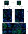
(a) Time- and stimulant-dependent internalization of overexpressed LGR5FL and LGR5Δ5 in HEK293T cells. The samples were stained with a FITC-coupled anti-Myc-Tag antibody (green) and counterstained (nuclei) with DAPI solution (blue). (b) Inhibition of clathrin- and caveolin-mediated endocytosis in LGR5FL-overexpressing and LGR5Δ5-overexpressing HEK293T cells. The cells were treated with inhibitors of clathrin-mediated endocytosis (MDC and PitStop2) and with an inhibitor of caveolin-mediated endocytosis (filipin III). (The scale bar represent 10µm). [20].
Figure 3
Figure 3
TOPFlash/FOPFlash reporter assay to measure Wnt pathway activity in modified HEK293T cells (a) overexpressing an empty vector (blue line), LGR5FL (black line), or LGR5Δ5 (red line). (b) siRNA-mediated knockdown of LGR4 and overexpression of an empty vector (light blue line), LGR5FL (black line), or LGR5Δ5 (red line). The control (dark blue line) corresponds to the empty vector control of (a).
Figure 4
Figure 4
Sanger sequencing of one CRISPR/Cas9 monoclonal cell line of HEK293T cells compared with the wild-type sequence. LGR4 exon 1 (−4 nucleotides) resulted in a stop codon in 629 and a 141 base pair mRNA encoding a truncated protein of 46 As), and LGR5 (−1 nucleotide) resulted in a stop codon in 661 and a 378 base pair mRNA encoding a truncated protein of 125 As. (Please also see the sequence of the rescue clones in Figure S1).
Figure 5
Figure 5
(a) TOP/FOP flash assay to measure Wnt pathway activity in modified HEK293T cells (empty vector (blue line), LGR5FL (black line), or LGR5Δ5 (red line) overexpression). (b) Screening of selected LGR4/5 CRISPR double-knockout clones via the TOPFlash/FOPFlash reporter assay after LGR5FL or LGRΔ5 rescue. Analysis of the chemiluminescence signals (luciferase activity) from transiently (pIRESpuro) LGR5FL- or LGR5Δ5-transfected cells with (+) and without (−) stimulation with 100 ng/mL Wnt3a and 100 ng/mL RSPO1. Control = knockout clones without transfection. Single-screen transfection was performed via ViaFect™. Clonal cell lines 1 and 3 were validated by sequencing analysis, whereas clone 2 was excluded.
Figure 6
Figure 6
Western blot analysis to detect full-length LGR5 (FL) and LGR5Δ5 (Δ5) expression via the lentiviral transduction of HEK LGR4/5 CRISPR/Cas9 double-knockout cells in comparison with the empty vector control (C). Full-length LGR5 (FL) and LGR5Δ5 (Δ5) were each tagged with a c-Myc tag (<1 kDa) or a YFP tag (27 kDa) for selection, visualization, and enrichment. The detection of LGR5 and LGR5Δ5 was carried out via a polyclonal anti-LGR5 antibody. ß actin was used as a loading control.
Figure 7
Figure 7
Migration of a HEK-LGR4/5 double-knockout clone with stable LGR5FL or LGR5Δ5 expression compared with the empty vector control cell line or unmodified HEK293T cell line via a scratch assay.
Figure 8
Figure 8
Radiosensitivity of the unmodified HEK293T cell line and the HEK-LGR4/5 double-knockout cell lines with stable LGR5FL or LGR5Δ5 expression or empty vector expression.
Figure 9
Figure 9
(a) Venn diagram of the HEK293T-LGR4/5 knockout cell lines (empty vector PLVX, LGR5FL, or LGR5Δ5 clones). (b) Volcano plot showing genes differentially expressed between the empty vector PLVX and LGR5Δ5 of the HEK293T-LGR4/5 knockout cell lines. (c) Heatmap of the expression of 15 genes related to LGR5Δ5 and LGR5FL overexpression (HEK293T-LGR4/5 knockout cell lines).
Figure 10
Figure 10
Western blot analysis of LGR5-regulated proteins in full-length LGR5 (FL)- and LGR5Δ5 (Δ5)-overexpressing HEK293T cells with single (LGR5) or double (LGR4/5) CRISPR Cas9 knockout with or without stimulation (Wnt3a and RSPO1) compared with control cells (PLVX) transfected with an empty vector. LGR5 and LGR5Δ5 were tagged with a YFP tag (27 kDa) for selection. ß actin was included as a loading control. Protein was isolated from adherent cells.
Figure 11
Figure 11
Postulated pathways associated with LGR5FL or LGR5Δ5 overexpression. LGR5FL activated the Wnt pathway, but LGR5Δ5 did not. LGR5FL overexpression is related to lupus erythematosus disease and fibrillar collagen regulation, whereas LGR5Δ5 overexpression is related to angiogenesis and fibrillar collagen regulation pathways. Our data also suggest that LGR5FL influences the level of genes associated with protein modification, transcription efficiency, and DNA accessibility adaptation in terms of epigenetic modifications. LGR5Δ5 seems to affect RSPO1 (the ligand of LGR5), an autoregulatory feedback loop activity of RSPO1. (Created with BioRender.com).

Similar articles

References

    1. de Lau W., Barker N., Low T.Y., Koo B.-K., Li V.S.W., Teunissen H., Kujala P., Haegebarth A., Peters P.J., van de Wetering M., et al. Lgr5 homologues associate with Wnt receptors and mediate R-spondin signalling. Nature. 2011;476:293–297. doi: 10.1038/nature10337. - DOI - PubMed
    1. Barker N., van Es J.H., Kuipers J., Kujala P., van den Born M., Cozijnsen M., Haegebarth A., Korving J., Begthel H., Peters P.J., et al. Identification of stem cells in small intestine and colon by marker gene Lgr5. Nature. 2007;449:1003–1007. doi: 10.1038/nature06196. - DOI - PubMed
    1. Leung C., Tan S.H., Barker N. Recent Advances in Lgr5+ Stem Cell Research. Trends Cell Biol. 2018;28:380–391. doi: 10.1016/j.tcb.2018.01.010. - DOI - PubMed
    1. Ghobakhloo S., Khoshhali M., Vatandoost N., Jafarpour S., Niazmand A., Nedaeinia R., Salehi R. Clinical Implications and Prognostic Value of Leucine-Rich G Protein-Coupled Receptor 5 Expression as A Cancer Stem Cell Marker in Malignancies: A Systematic Review and Meta-Analysis. Cell J. 2024;26:1–12. doi: 10.22074/CELLJ.2023.2010157.1396. - DOI - PMC - PubMed
    1. Barker N., Clevers H. Leucine-rich repeat-containing G-protein-coupled receptors as markers of adult stem cells. Gastroenterology. 2010;138:1681–1696. doi: 10.1053/j.gastro.2010.03.002. - DOI - PubMed

MeSH terms

LinkOut - more resources