. 2024 Dec 15;25(24):13434.
doi: 10.3390/ijms252413434.

Preliminary Study on the Positive Expression Regulation of Alpha2-Macroglobulin in the Testicular Tissue of Male Mice by Environmental Estrogens

Affiliations

Preliminary Study on the Positive Expression Regulation of Alpha2-Macroglobulin in the Testicular Tissue of Male Mice by Environmental Estrogens

Hong-Mei Li et al. Int J Mol Sci. .

Abstract

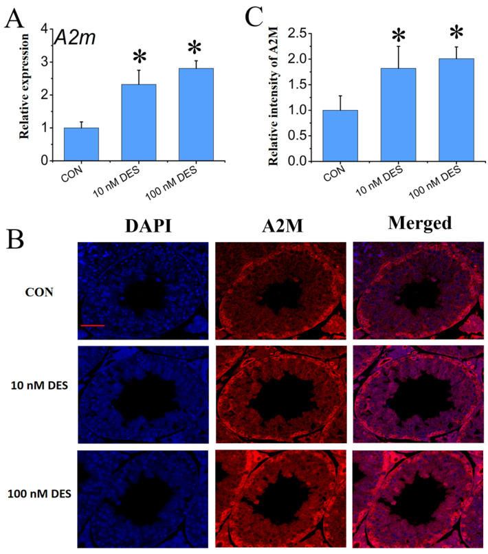
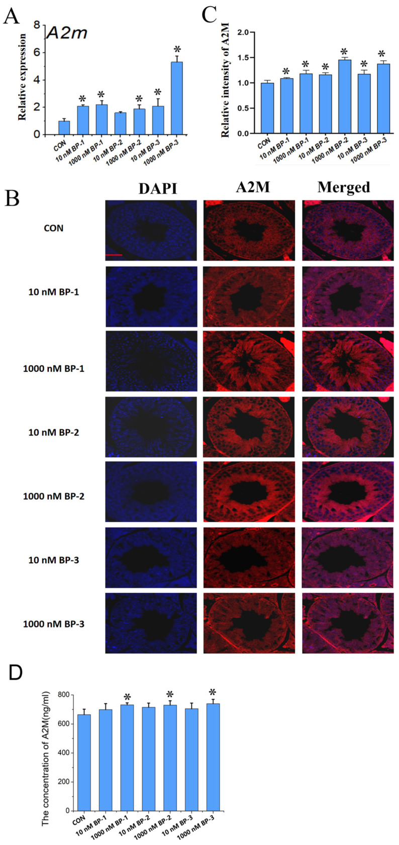
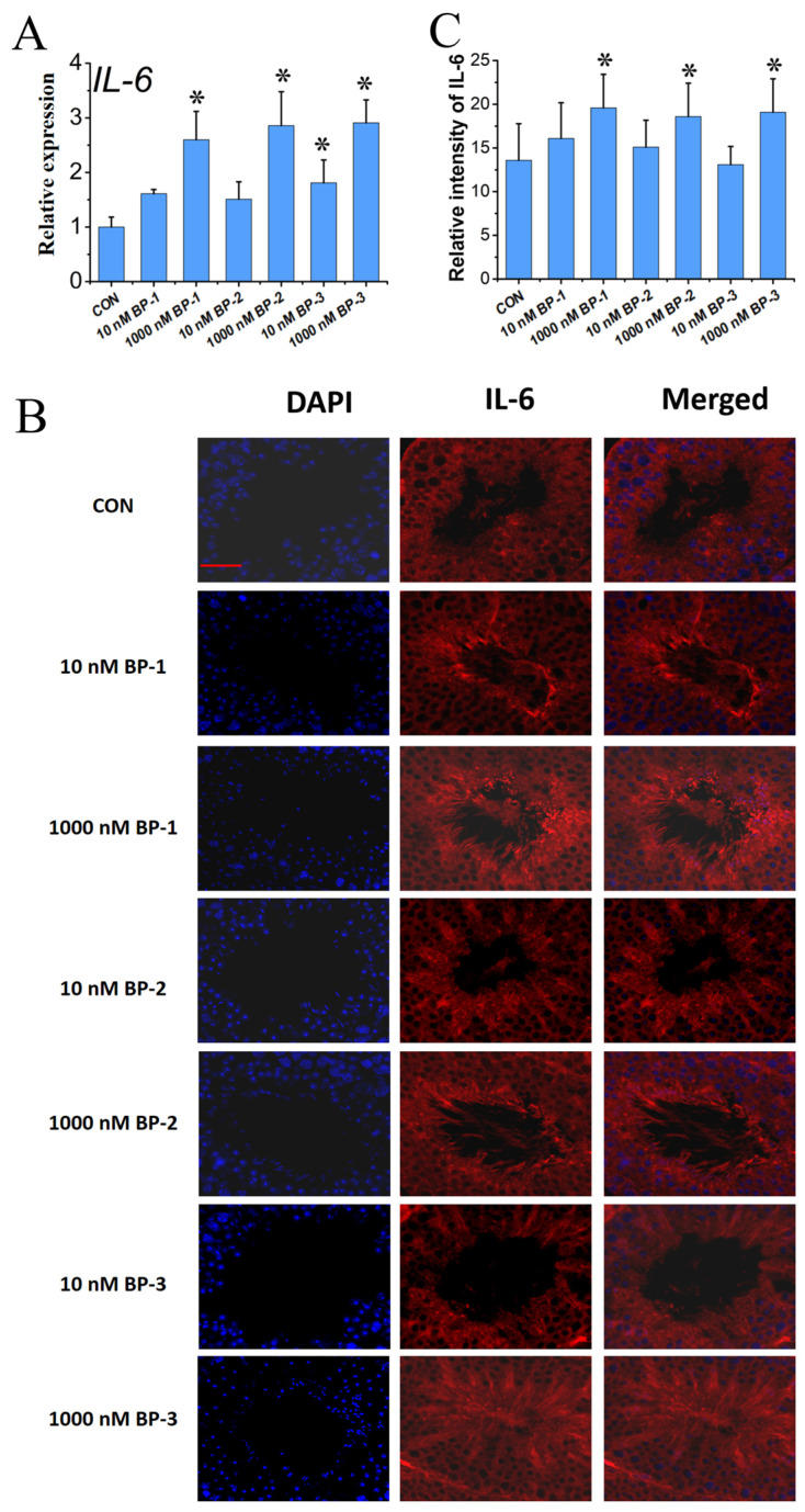
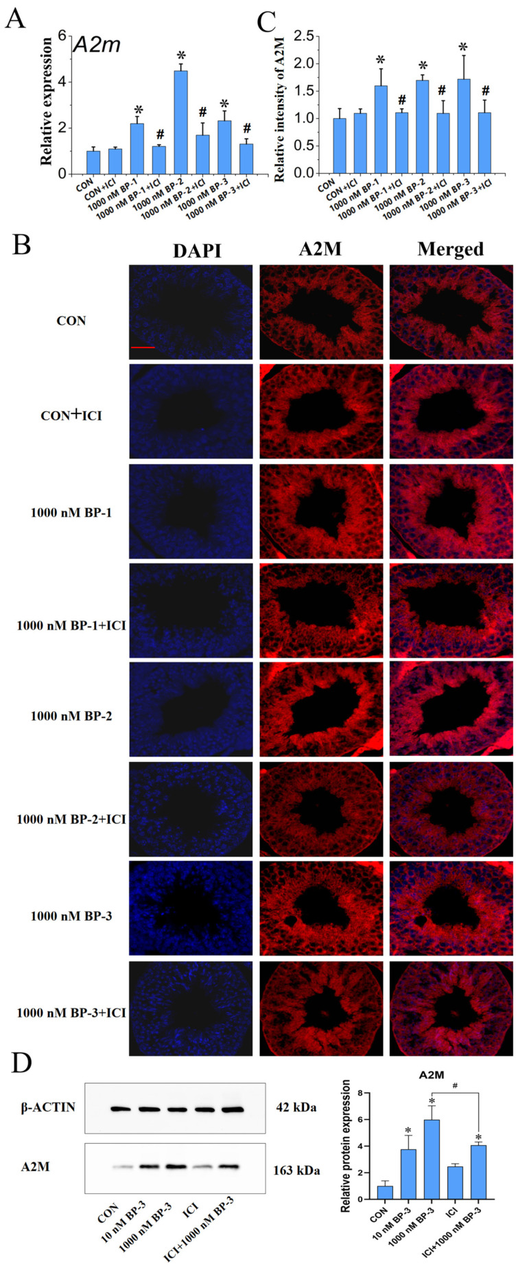
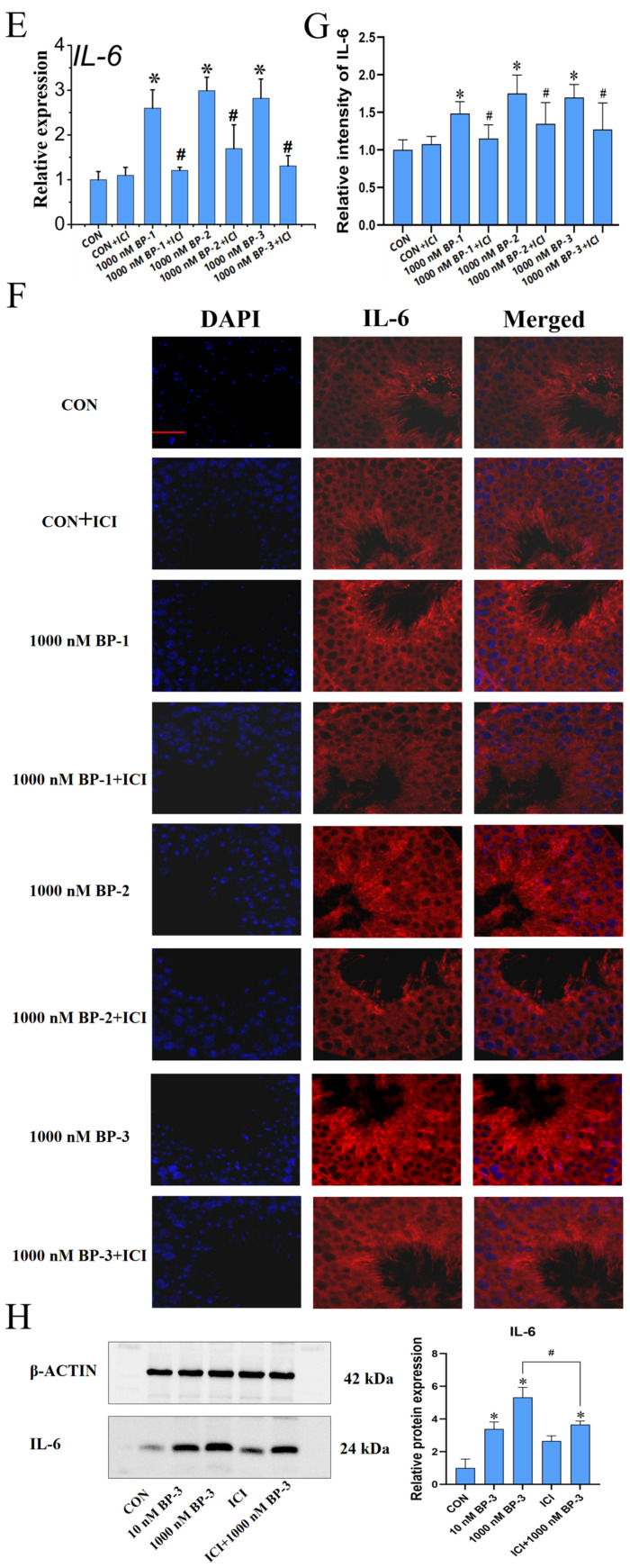
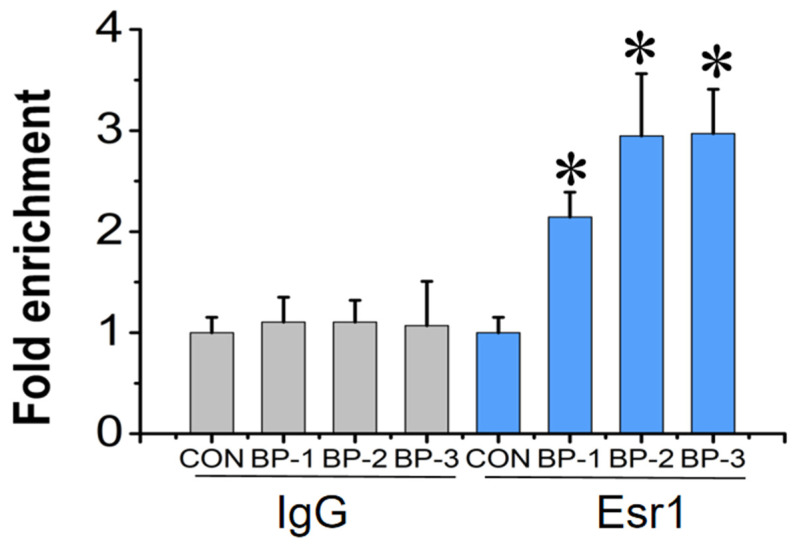
The male reproductive impairment caused by environmental estrogens (EEs) stands as a pivotal research area in environmental toxicology. Alpha2-macroglobulin (A2M) emerges as a promising molecule capable of counteracting oxidative stress induced by EEs. This study conducted exposure experiments spanning PND1 to PND56 employing ICR mice, aiming to delve into the expression patterns of A2M and its modulated IL-6 in the testicular tissue of mice subsequent to diethylstilbestrol (DES) and benzophenone (BP) exposure, while elucidating the pivotal role of ERs in this intricate process. Our findings revealed that upon DES exposure (10 and 100 nM), there was a pronounced upregulation of A2M (mRNA and in situ protein levels) in mouse testicular tissue. Similarly, exposure to BPs (BP-1, BP-2, and BP-3, each at 10 and 1000 nM) exhibited comparable effects and increasing A2M levels in serum. Notably, BP exposure also caused an elevation in IL-6 levels (which could be directly regulated by A2M) within testicular tissue (mRNA and in situ protein). Remarkably, the specific estrogen receptor antagonist ICI 182780 (0.5 mg/kg/day) was effective in reversing the upregulation of both A2M and IL-6 induced by BP exposure. Significantly, the results of theoretical prediction of the potential ERE site in the A2m gene promoter region and ChIP-qPCR experiment provide essential and strong evidence for the key conclusion that A2m is the target gene of ER. Taken together, our study highlights EEs' ability to regulate A2M expression in the male reproductive system via the ER signaling pathway. This vital insight deepens our understanding of molecular mechanisms protecting against oxidative stress caused by EEs.

Keywords: alpha2-macroglobulin (A2M); environmental estrogens (EEs); estrogen receptor (ER); interleukin-6 (IL-6); male reproductive system.


Conflict of interest statement

The authors declare no conflicts of interest.

Figures

Figure 1
Figure 1
The effect of DES exposure on the expression level of A2M in mouse testes (PND1–PND56) (A). Relative mRNA expression level (B). In situ protein expression level (IF assay) (C). Quantitative analysis of IF assay results. Scale bar: 50 μm. Data are expressed as mean ± SD. * Indicates significant differences between BP groups and the control group (p < 0.05).
Figure 2
Figure 2
The effect of BP exposure on the expression level of A2M in mouse testes (PND1–PND56) (A). Relative mRNA expression level (qPCR). (B). In situ protein expression level (IF assay). (C). Quantitative analysis of IF assay results. (D). The concentration of A2M in serum. Scale bar: 50 μm. Data are expressed as mean ± SD. * Indicates significant differences between BP groups and the control group (p < 0.05).
Figure 3
Figure 3
The effects of BP exposure on the expression level of IL-6 in mouse testes (PND1–PND56) (A). Relative mRNA expression level (qPCR). (B). In situ protein expression level (IF assay). (C). Quantitative analysis of IF assay results. Scale bar: 50 μm. Data are expressed as mean ± SD. * Indicates significant differences between BP groups and the control group (p < 0.05).
Figure 4
Figure 4
The effects of ICI intervention on the upregulation of A2M and IL-6 in testicular tissue of mice exposed to BPs. (AC) Relative mRNA expression level (qPCR), in situ protein expression level (IF assay), and the quantitative analysis of IF assay results of A2M, respectively. (DF) Relative mRNA expression level (qPCR), in situ protein expression level (IF assay), and the quantitative analysis of IF assay results of IL-6, respectively. (G,H) Protein quantitative expression levels (WB assay), and the quantitative analysis of WB assay results of A2M and IL-6, respectively. Scale bar: 50 μm. Data are expressed as mean ± SD. * Indicates significant differences between BP groups and the control group (p < 0.05). # Indicates significant difference between BPs groups and the corresponding ICI 182780 intervention groups (p < 0.05).
Figure 4
Figure 4
The effects of ICI intervention on the upregulation of A2M and IL-6 in testicular tissue of mice exposed to BPs. (AC) Relative mRNA expression level (qPCR), in situ protein expression level (IF assay), and the quantitative analysis of IF assay results of A2M, respectively. (DF) Relative mRNA expression level (qPCR), in situ protein expression level (IF assay), and the quantitative analysis of IF assay results of IL-6, respectively. (G,H) Protein quantitative expression levels (WB assay), and the quantitative analysis of WB assay results of A2M and IL-6, respectively. Scale bar: 50 μm. Data are expressed as mean ± SD. * Indicates significant differences between BP groups and the control group (p < 0.05). # Indicates significant difference between BPs groups and the corresponding ICI 182780 intervention groups (p < 0.05).
Figure 5
Figure 5
Quantitative enrichment of A2m after chromatin immunoprecipitation (ChIP) with Esr1antibody and immunoglobulin G (IgG) in mouse testes following postnatal exposure to 1000 nM BPs (BP-1, BP-2, and BP-3) for 56 days. Data were expressed as mean ± SD. * Indicates significant difference between BP groups and the control group (p < 0.05).

Similar articles

References

    1. Ma Y., Shen W., Tang T., Li Z., Dai R. Environmental estrogens in surface water and their interaction with microalgae: A review. Pt 1Sci. Total Environ. 2022;807:150637. doi: 10.1016/j.scitotenv.2021.150637. - DOI - PubMed
    1. Matta M.K., Zusterzeel R., Pilli N.R., Patel V., Volpe D.A., Florian J., Oh L., Bashaw E., Zineh I., Sanabria C., et al. Effect of sunscreen application under maximal use conditions on plasma concentration of sunscreen active ingredients: A randomized clinical trial. JAMA. 2019;321:2082–2091. doi: 10.1001/jama.2019.5586. - DOI - PMC - PubMed
    1. Adeel M., Song X., Wang Y., Francis D., Yang Y. Environmental impact of estrogens on human, animal and plant life: A critical review. Environ. Int. 2017;99:107–119. doi: 10.1016/j.envint.2016.12.010. - DOI - PubMed
    1. Chen P., Li B., Ou-Yang L. Role of estrogen receptors in health and disease. Front. Endocrinol. 2022;13:839005. doi: 10.3389/fendo.2022.839005. - DOI - PMC - PubMed
    1. Newbold R.R. Lessons learned from perinatal exposure to diethylstilbestrol. Toxicol. Appl. Pharmacol. 2004;199:142–150. doi: 10.1016/j.taap.2003.11.033. - DOI - PubMed

LinkOut - more resources