. 2024 Dec 15;25(24):13439.
doi: 10.3390/ijms252413439.

Chiral Amino Acids Mediate Mitochondria-Dependent Apoptosis of Human Proximal Tubular Epithelial Cells Under Oxidative Stress

Affiliations

Chiral Amino Acids Mediate Mitochondria-Dependent Apoptosis of Human Proximal Tubular Epithelial Cells Under Oxidative Stress

Ying Lu et al. Int J Mol Sci. .

Abstract

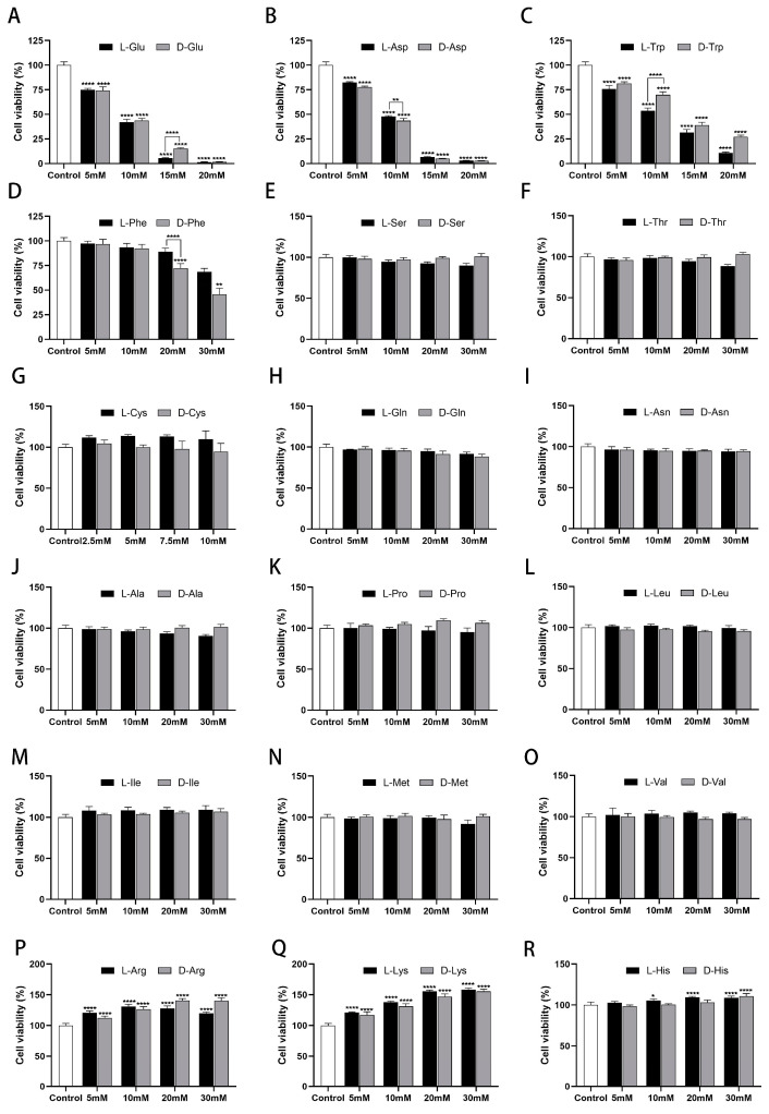
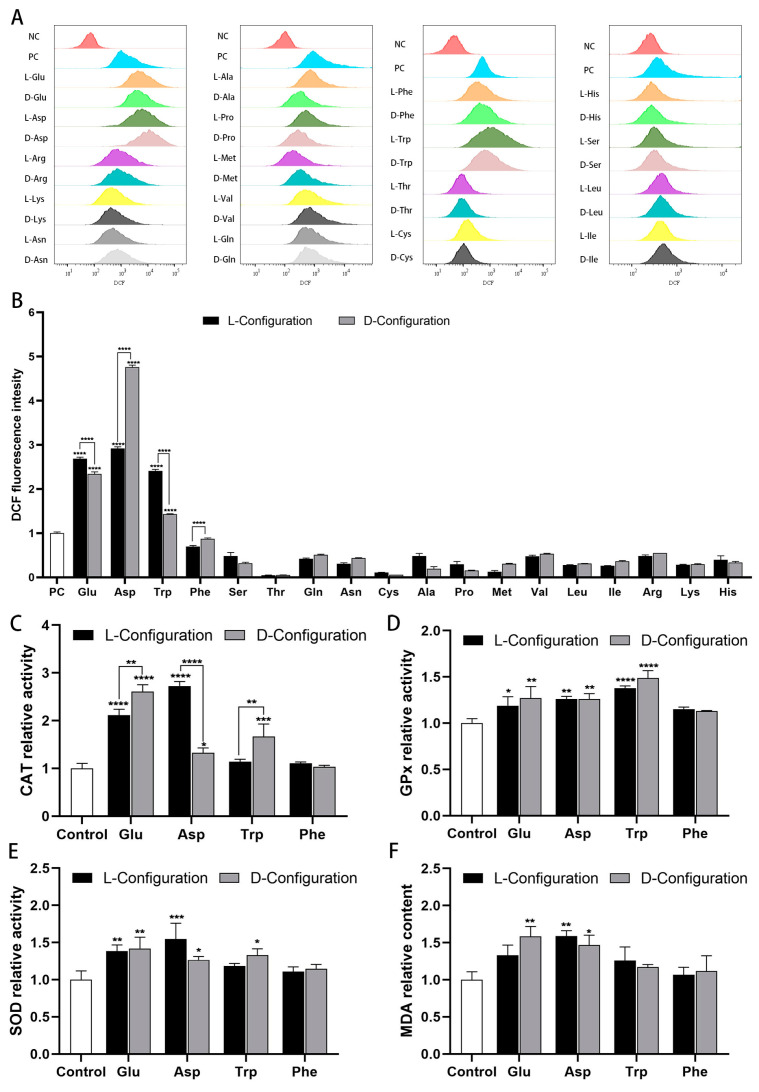
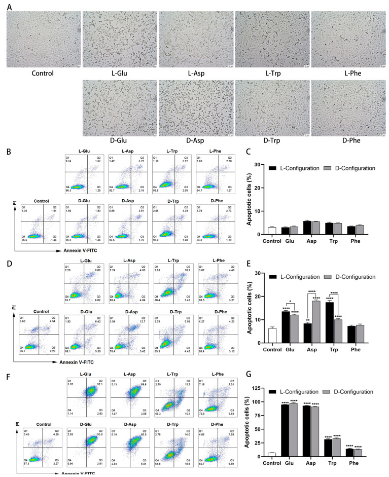
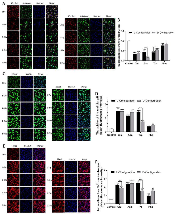
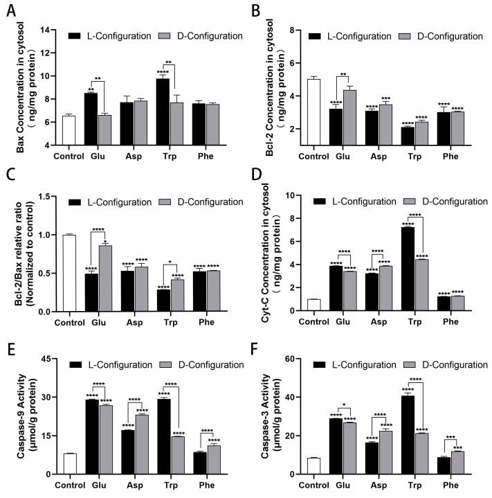
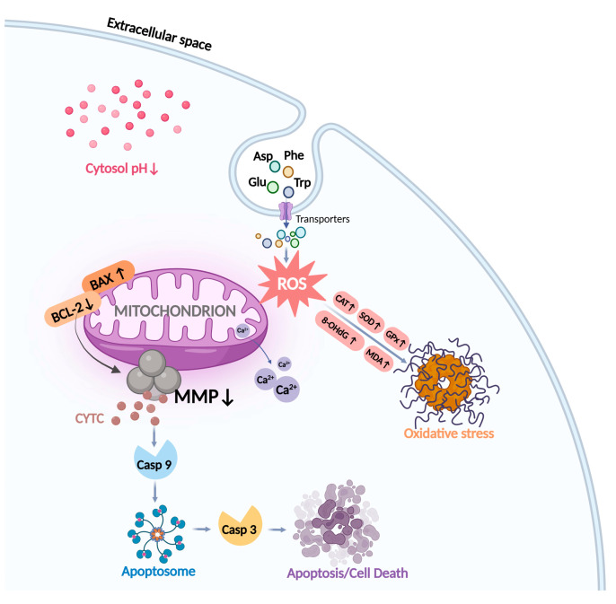
Amino acids are the basic structural units of life, and their intake levels affect disease and health. In the case of renal disease, alterations in amino acid metabolism can be used not only as a clinical indicator of renal disease but also as a therapeutic strategy. However, the biological roles and molecular mechanisms of natural chiral amino acids in human proximal tubular epithelial cells (HK-2) remain unclear. In this study, cell viability assays revealed that chiral acidic amino acids (Glu and Asp) and aromatic amino acids (Trp and Phe) inhibited cell growth. The molecular mechanisms indicated that cell growth was closely related to ROS levels. Specifically, chiral Glu, Asp, Trp, and Phe induced oxidative stress and mitochondria-dependent apoptosis in HK-2 cells. This was manifested by elevated levels of intracellular ROS, 8-OHdG, and MDA, increased activities of antioxidant enzymes CAT, SOD, and GPx, decreased mitochondrial membrane potential, increased cytoplasmic Ca2+ concentration, and cell acidification. The expression levels of apoptosis-related molecules Caspase-9, Caspase-3, Cyt-C, and Bax were increased, and the expression level of anti-apoptotic molecule Bcl-2 was decreased. Moreover, L-Glu, D-Asp, L-Trp, and D-Phe exhibited a more pronounced inhibition of cell growth and elicited more substantial alterations in gene expression compared to the other configurations.

Keywords: HK-2 cells; ROS; apoptosis; chiral amino acids; mitochondria-dependent; oxidative stress.


Conflict of interest statement

The authors declare no conflicts of interest.

Figures

Figure 1
Figure 1
Effects of chiral amino acids on HK-2 cells’ viability. (A,B) Acidic amino acid (Glu, Asp). (C,D) Aromatic amino acids (Trp, Phe). (EO) Neutral amino acids. (PR) Basic amino acids. (Statistical significance relative to control was marked with * p < 0.05, ** p < 0.01, or **** p < 0.0001).
Figure 2
Figure 2
Oxidative stress induced by 10 mM of chiral amino acids acting on HK-2 cells for 24 h. (A) Fluorescence distribution of DCFH-DA probe, where negative control (NC) was without amino acids and ROS-up treatment, and positive control (PC) was with ROS-up treatment. (B) Mean fluorescence intensity of the DCFH-DA probe, where all groups were subtracted from the autofluorescence value of the negative control. Relative activity of (C) catalase, (D) glutathione peroxidase, (E) superoxide dismutase, and (F) lipid peroxidation. (Statistical significance relative to control was marked with * p < 0.05, ** p < 0.01, *** p < 0.001, or **** p < 0.0001).
Figure 3
Figure 3
Morphological changes and apoptosis induced by chiral amino acids in HK-2 cells under oxidative stress. (A) Cell morphology after 15 mM of chiral amino acids acting on HK-2 cells for 24 h, observed with an inverted microscope at a magnification of 10×. Scale bars: 50 µm. Double staining of apoptotic cells and quantification of apoptosis rate after (B,C) 10 mM, (D,E) 15 mM, and (F,G) 20 mM of chiral amino acids acting on HK-2 cells for 24 h. (Statistical significance relative to control was marked with * p < 0.05, ** p < 0.01, or **** p < 0.0001).
Figure 4
Figure 4
Mitochondrial dysfunction induced after 15 mM of chiral amino acids acting on HK-2 cells for 24 h. (A) Representative confocal images of HK-2 cells stained with JC-1 (red and green) and Hoechst 33342 (blue). Scale bars: 20 µm. (B) Quantification of the level of MMP. (C) Representative confocal images of HK-2 cells stained with BCECF (green) and Hoechst 33342 (blue). Scale bars: 20 µm. (D) Quantification of the extent of cell acidification. (E) Representative confocal images of HK-2 cells stained with Rhod (red) and Hoechst 33342 (blue). Scale bars: 20 µm. (F) Quantification of the concentration of intracellular free Ca2+. (Statistical significance relative to control was marked with * p < 0.05, ** p < 0.01, *** p < 0.001, or **** p < 0.0001).
Figure 5
Figure 5
Variable expressions of mitochondria-dependent apoptosis-related proteins induced after 15 mM of chiral amino acids acting on HK-2 cells for 24 h. (A,B) Bax and Bcl-2 protein concentrations. (C) Relative ratio of Bcl-2/Bax protein after normalization. (D) Cyt-C protein content. (E,F) Caspase-9 and Caspase-3 activity. (Statistical significance relative to control was marked with * p < 0.05, ** p < 0.01, *** p < 0.001, or **** p < 0.0001).
Figure 6
Figure 6
Diagram of the molecular mechanism of chiral amino acid action in HK-2 cells. After exposure to Glu, Asp, Trp, and Phe, the mitochondrial ROS levels in HK-2 cells increased, inducing oxidative damage to DNA and lipids. Redox homeostasis is regulated by elevating antioxidant enzyme levels, and an imbalance in this homeostasis leads to oxidative stress. Meanwhile, decreased cellular MMP, mitochondrial Ca2+ release, and cellular acidification mediated altered mitochondrial function, which further induced apoptosis through the action of the Bcl-2 family, Cyt-C, and Caspase family protein pathways.

Similar articles

References

    1. Tabe Y., Lorenzi P.L., Konopleva M. Amino acid metabolism in hematologic malignancies and the era of targeted therapy. Blood. 2019;134:1014–1023. doi: 10.1182/blood.2019001034. - DOI - PMC - PubMed
    1. Massey K.A., Blakeslee C.H., Pitkow H.S. A review of physiological and metabolic effects of essential amino acids. Amino Acids. 1998;14:271–300. doi: 10.1007/BF01318848. - DOI - PubMed
    1. Giri C., Goesmann F., Meinert C., Evans A.C., Meierhenrich U.J. Synthesis and Chirality of Amino Acids Under Interstellar Conditions. Topics Curr. Chem. 2013;333:57–61. doi: 10.1007/128_2012_367. - DOI - PubMed
    1. Meinert C., Garcia A.D., Topin J., Jones N.C., Diekmann M., Berger R., Nahon L., Hoffmann S.V., Meierhenrich U.J. Amino acid gas phase circular dichroism and implications for the origin of biomolecular asymmetry. Nat. Commun. 2022;13:502. doi: 10.1038/s41467-022-28184-0. - DOI - PMC - PubMed
    1. Marcone G.L., Rosini E., Crespi E., Pollegioni L. D-amino acids in foods. Appl. Microbiol. Biot. 2020;104:555–574. doi: 10.1007/s00253-019-10264-9. - DOI - PubMed

MeSH terms

LinkOut - more resources