. 2024 Dec 16;25(24):13482.
doi: 10.3390/ijms252413482.

Molecular Insights into Structural Dynamics and Binding Interactions of Selected Inhibitors Targeting SARS-CoV-2 Main Protease

Affiliations

Molecular Insights into Structural Dynamics and Binding Interactions of Selected Inhibitors Targeting SARS-CoV-2 Main Protease

Yuanyuan Wang et al. Int J Mol Sci. .

Abstract

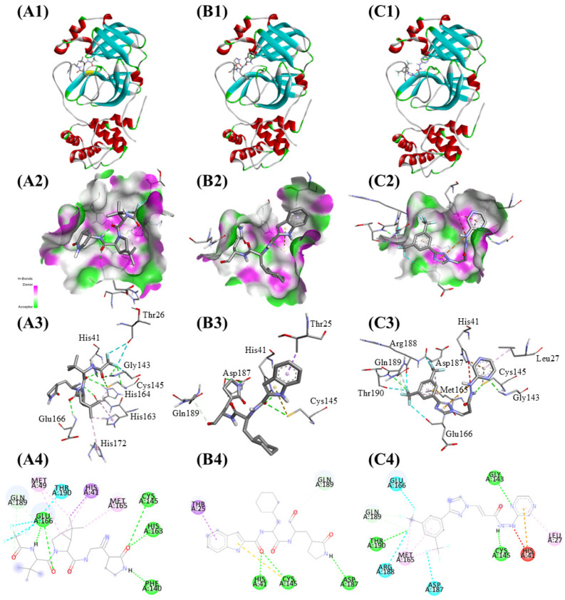
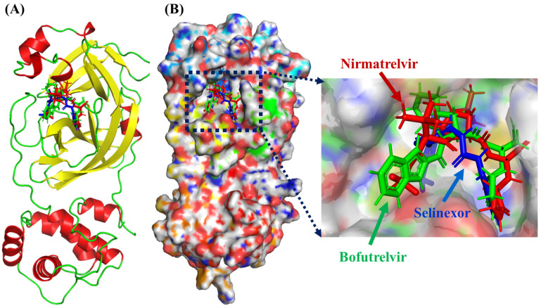
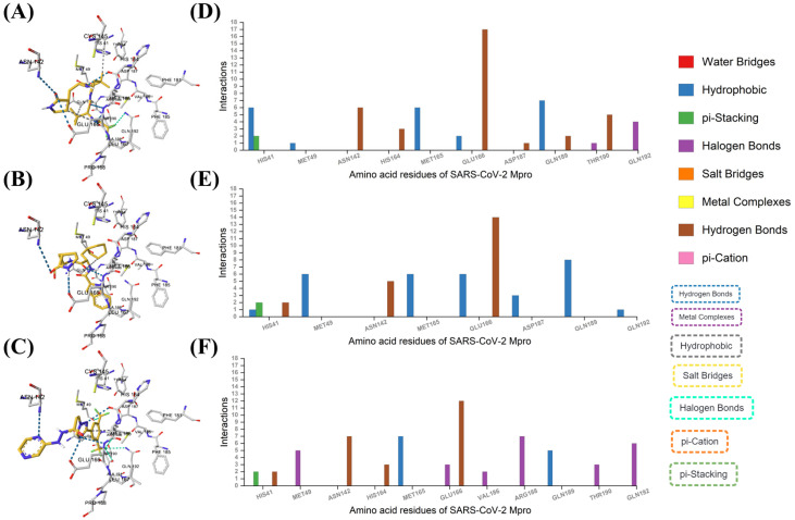
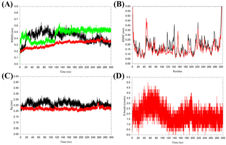
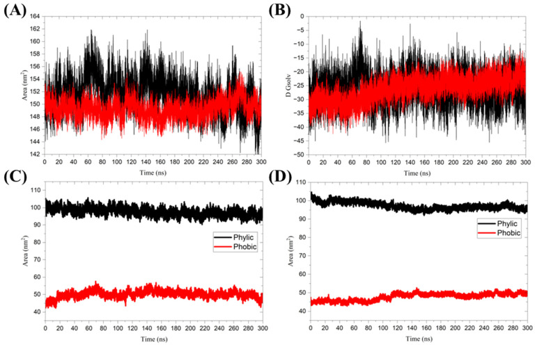
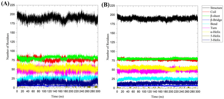
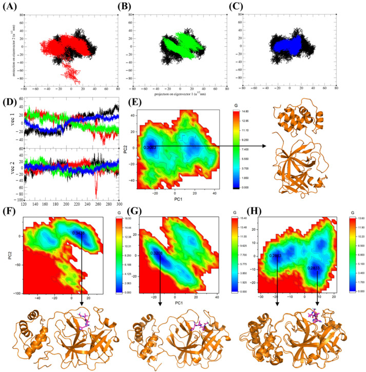
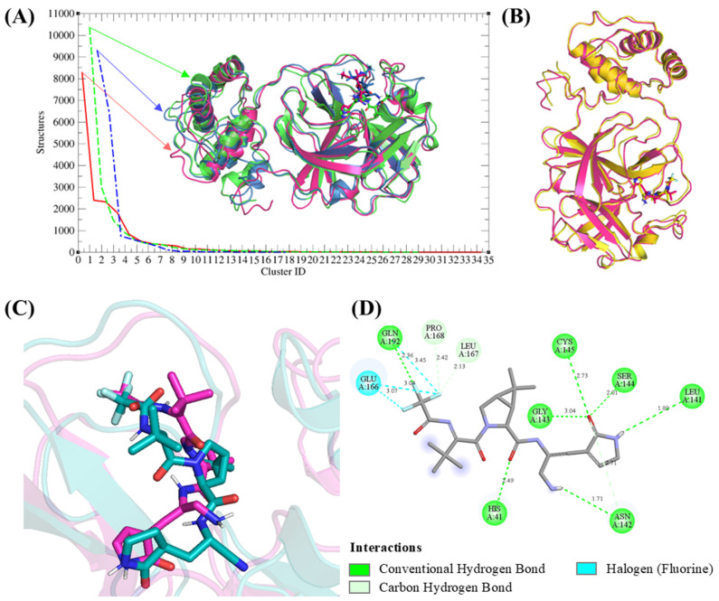
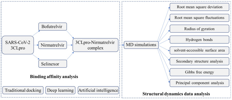
The SARS-CoV-2 main protease (Mpro, also known as 3CLpro) is a key target for antiviral therapy due to its critical role in viral replication and maturation. This study investigated the inhibitory effects of Bofutrelvir, Nirmatrelvir, and Selinexor on 3CLpro through molecular docking, molecular dynamics (MD) simulations, and free energy calculations. Nirmatrelvir exhibited the strongest binding affinity across docking tools (AutoDock Vina: -8.3 kcal/mol; DiffDock: -7.75 kcal/mol; DynamicBound: 7.59 to 7.89 kcal/mol), outperforming Selinexor and Bofutrelvir. Triplicate 300 ns MD simulations revealed that the Nirmatrelvir-3CLpro complex displayed high conformational stability, reduced root mean square deviation (RMSD), and a modest decrease in solvent-accessible surface area (SASA), indicating enhanced structural rigidity. Gibbs free energy analysis highlighted greater flexibility in unbound 3CLpro, stabilized by Nirmatrelvir binding, supported by stable hydrogen bonds. MolProphet prediction tools, targeting the Cys145 residue, confirmed that Nirmatrelvir exhibited the strongest binding, forming multiple hydrophobic, hydrogen, and π-stacking interactions with key residues, and had the lowest predicted IC50/EC50 (9.18 × 10⁻⁸ mol/L), indicating its superior potency. Bofutrelvir and Selinexor showed weaker interactions and higher IC50/EC50 values. MM/PBSA analysis calculated a binding free energy of -100.664 ± 0.691 kJ/mol for the Nirmatrelvir-3CLpro complex, further supporting its stability and binding potency. These results underscore Nirmatrelvir's potential as a promising therapeutic agent for SARS-CoV-2 and provide novel insights into dynamic stabilizing interactions through AI-based docking and long-term MD simulations.

Keywords: Bofutrelvir; Paxlovid; SARS-CoV-2; Selinexor; artificial intelligence; molecular interactions.


Conflict of interest statement

The authors declare no conflicts of interest.

Figures

Figure 1
Figure 1
(A) Crystal structure of SARS-CoV-2 3CLpro (PDB: 1P9S) showing two monomers (monomer A in red and monomer B in green). (B) Crystal structure of the SARS-CoV-2 3CLpro monomer (PDB: 7ALH) showing the three domains.
Figure 2
Figure 2
Binding interactions of the three compounds with SARS-CoV-2 3CLpro: (A) Nirmatrelvir, (B) Selinexor, and (C) Bofutrelvir. (1) Active site view showing drug binding. (2) Surface view highlighting hydrogen bond donors and acceptors. (3) Atom-level view of the interactions between the ligand and key active-site residues. (4) 2D interaction map including hydrogen bonds (green dashed lines), hydrophobic interactions (purple dashed lines), and unfavorable contacts (red dashed lines).
Figure 3
Figure 3
Deep equivariant generative model sampling. (A) The dynamic docking investigation of Nirmatrelvir (red), Bofutrelvir (green), and Selinexor (blue) into the active pocket of SARS-CoV-2 Mpro. In the protein structure, α-helices are shown in red, β-sheets in yellow, and loop regions in green. (B) Protein surface view showing the dynamic poses of these ligands.
Figure 4
Figure 4
AI-based molecular interactions. The binding modes of (A) Nirmatrelvir, (B) Bofutrelvir, and (C) Selinexor with SARS-CoV-2 Mpro. Analysis of the interactions of SARS-CoV-2 Mpro residues with (D) Nirmatrelvir, (E) Bofutrelvir, and (F) Selinexor.
Figure 5
Figure 5
Molecular dynamics simulations of SARS-CoV-2 3CL protease in its Nirmatrelvir-unbound and Nirmatrelvir-bound forms. (A) Root mean square deviation (RMSD), (B) Root mean square fluctuations (RMSF), (C) Radius of gyration (Rg), and (D) Hydrogen bond analysis between Nirmatrelvir and 3CL protease. The color black represents 3CL protease alone, while red indicates the 3CL protease in the Nirmatrelvir-3CL protease complex, and green represents the ligand in the Nirmatrelvir-3CL protease complex. The presented charts are the average representation of the triplicate MD simulation runs for the Nirmatrelvir-3CL protease complex.
Figure 6
Figure 6
(A) Solvent-accessible surface area, and (B) Free energy of solvation. The color black represents 3CL protease alone, while red indicates the 3CL protease in the Nirmatrelvir-3CL protease complex. The SASA was further divided into hydrophobic and hydrophilic regions for (C) 3CL protease alone and (D) 3CL protease in the Nirmatrelvir-3CL protease complex. The presented charts are the average representation of the triplicate MD simulation runs for the Nirmatrelvir-3CL protease complex.
Figure 7
Figure 7
Secondary structure analysis during the 300 ns MD simulation for (A) unbound 3CL protease and (B) 3CL protease in the Nirmatrelvir-3CL protease complex. The presented charts are the average representation of the triplicate MD simulation runs for the Nirmatrelvir-3CL protease complex. Structure = α-helix + β-sheet + β-bridge + Turn.
Figure 8
Figure 8
Principal component analysis (PCA) trajectory projections of 3CLpro along eigenvector 1 (PC1) and eigenvector 2 (PC2), comparing the Nirmatrelvir-unbound form and Nirmatrelvir-bound complexes. Trajectory projections for (A) replicate 1, (B) replicate 2, and (C) replicate 3. (D) Eigenvector analysis of the 3CLpro in its Nirmatrelvir-unbound form and Nirmatrelvir-bound complexes. The black color represents the Nirmatrelvir-unbound 3CL protease, while red, green, and blue correspond to the complexed 3CL protease in replicates 1, 2, and 3, respectively. (E) Gibbs free energy (GFE) landscape and the representative structure with the lowest free energy of the Nirmatrelvir-unbound 3CL protease, and (FH) GFE landscapes and the representative structures with the lowest free energy of the 3CL protease in the Nirmatrelvir-3CL protease complex for replicates 1, 2, and 3. The numbers 0.3083, 0.3438, 0.3208, and 0.2833 represent the free energy values (in kcal/mol) of the minima energy basins, indicating the most stable conformational states within the energy landscape.
Figure 9
Figure 9
Clustering and binding analysis of the Nirmatrelvir-3CL protease complex. (A) Cluster analysis identified 35, 29, and 17 clusters for replicas 1, 2, and 3, respectively. (B) Superimposition of the representative structure of replica 1 (magenta) with its lowest-energy structure (yellow), showing close alignment. (C) Superimposition of Nirmatrelvir in replica 1’s representative structure (magenta) with its docked pose (teal), revealing minimal deviations. (D) 2D interaction analysis of Nirmatrelvir with 3CL protease based on the representative structure of replica 1.
Figure 10
Figure 10
The flowchart outlining the analysis of binding affinity and structural dynamics of SARS-CoV-2 3CLpro and its inhibitors.

Similar articles

Cited by

References

    1. Singh M., Jayant K., Singh D., Bhutani S., Poddar N.K., Chaudhary A.A., Khan S.U., Adnan M., Siddiqui A.J., Hassan M.I., et al. Withania somnifera (L.) Dunal (Ashwagandha) for the possible therapeutics and clinical management of SARS-CoV-2 infection: Plant-based drug discovery and targeted therapy. Front. Cell Infect. Microbiol. 2022;12:933824. doi: 10.3389/fcimb.2022.933824. - DOI - PMC - PubMed
    1. Ciotti M., Angeletti S., Minieri M., Giovannetti M., Benvenuto D., Pascarella S., Sagnelli C., Bianchi M., Bernardini S., Ciccozzi M. COVID-19 Outbreak: An Overview. Chemotherapy. 2019;64:215–223. doi: 10.1159/000507423. - DOI - PMC - PubMed
    1. Fagbule O.F. 2019 Novel Coronavirus. Ann. Ibd. Postgrad. Med. 2019;17:108–110. - PMC - PubMed
    1. Liu S.L. New virus in China requires international control effort. Nature. 2020;577:472. doi: 10.1038/d41586-020-00135-z. - DOI - PubMed
    1. Jee Y. WHO International Health Regulations Emergency Committee for the COVID-19 outbreak. Epidemiol. Health. 2020;42:e2020013. doi: 10.4178/epih.e2020013. - DOI - PMC - PubMed

MeSH terms

Grants and funding

LinkOut - more resources