. 2024 Dec 17;25(24):13524.
doi: 10.3390/ijms252413524.

Itchy E3 Ubiquitin Ligase-Mediated Ubiquitination of Ferritin Light Chain Contributes to Endothelial Ferroptosis in Atherosclerosis

Affiliations

Itchy E3 Ubiquitin Ligase-Mediated Ubiquitination of Ferritin Light Chain Contributes to Endothelial Ferroptosis in Atherosclerosis

Yi Zeng et al. Int J Mol Sci. .

Abstract

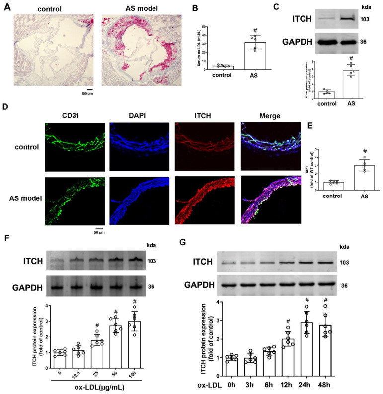
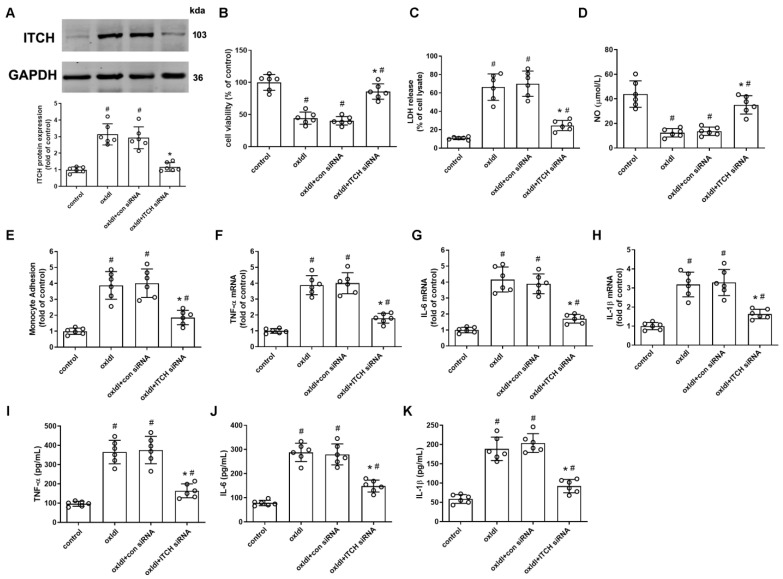
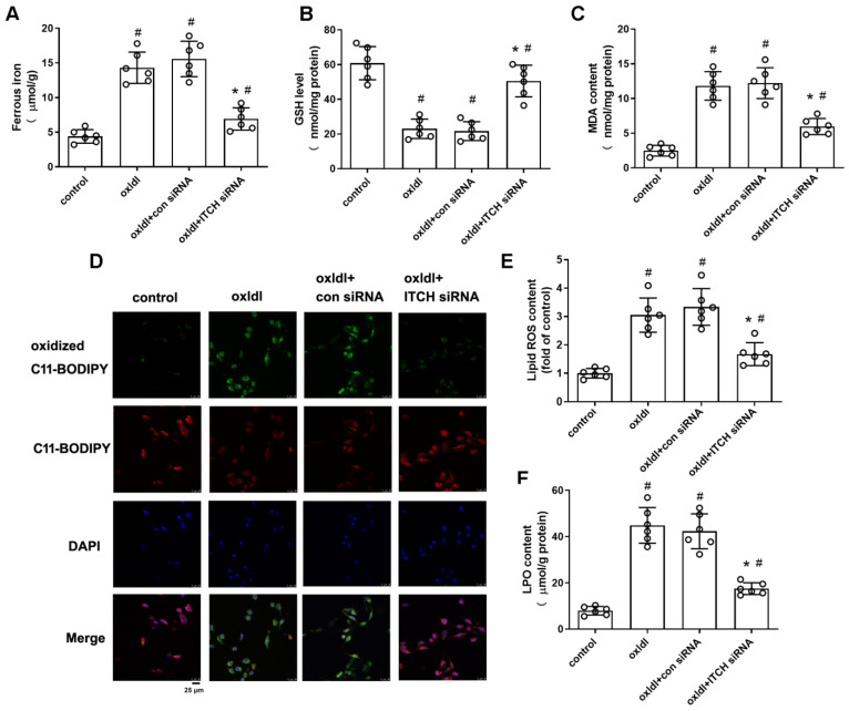
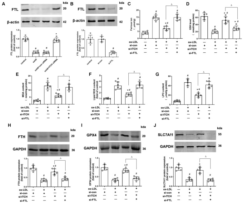
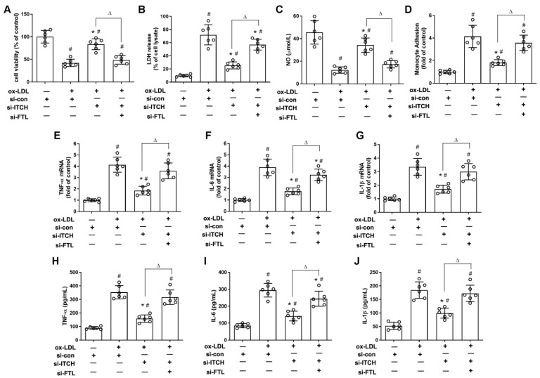
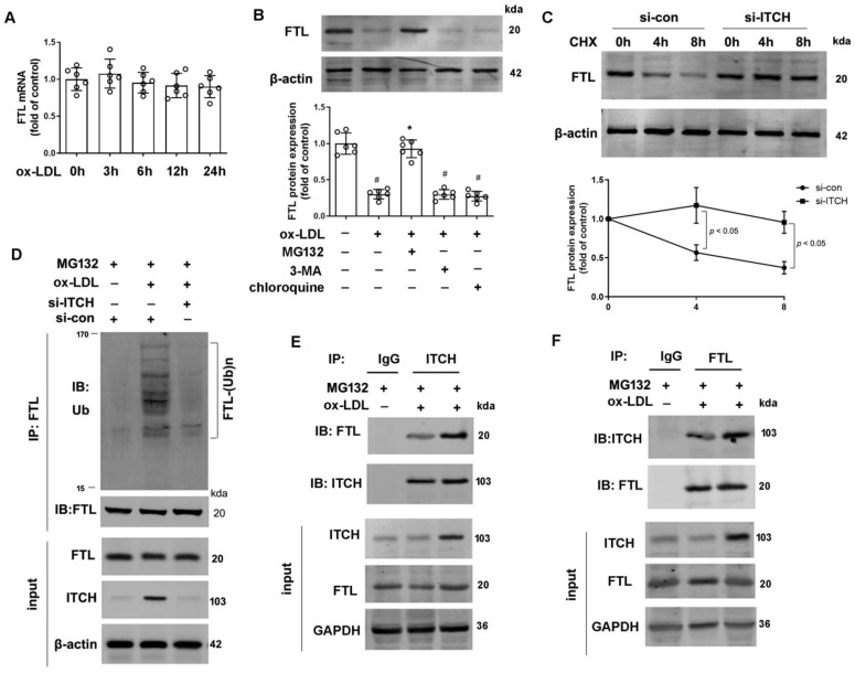
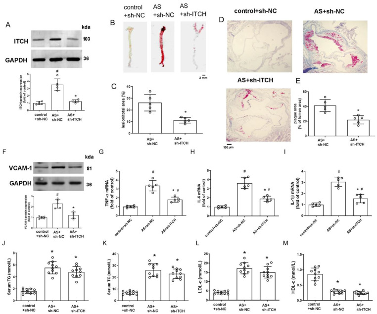
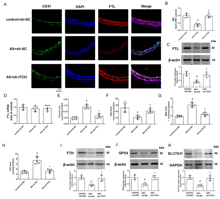
This research seeks to investigate the function and fundamental mechanisms of Itchy E3 ubiquitin ligase (ITCH), a HECT (homologous to E6AP carboxyl terminus)-type E3 ubiquitin ligase, in endothelial ferroptosis, particularly in the context of atherosclerosis, which has been underexplored. The levels of ITCH protein in the aortas of mice with atherosclerosis were analyzed. Constructs for ITCH RNA interference were generated and introduced into human aortic endothelial cells (HAECs). The findings indicated that ITCH protein expression was elevated in atherosclerotic mice and HAECs exposed to oxidized low-density lipoprotein (ox-LDL). ITCH downregulation significantly mitigated ox-LDL-induced endothelial injury and dysfunction. Reducing ITCH expression inhibited ox-LDL-induced endothelial ferroptosis. This study also revealed that ITCH mediates ox-LDL-induced ubiquitin-dependent degradation of ferritin light chain (FTL) in HAECs. The protective impact of ITCH knockdown against ox-LDL-induced ferroptosis and endothelial injury was reversed by FTL siRNA. Additionally, in vivo experiments showed that inhibiting ITCH reduced atherosclerosis progression and reversed ferroptosis in the aorta, with an associated increase in FTL protein expression in the aortas of mice. This study demonstrates that ITCH interacts with and regulates the stability of the FTL protein via the ubiquitin-proteasome system, contributing to ox-LDL-induced ferroptosis and endothelial cell dysfunction. Targeting components of the ITCH-FTL pathway holds potential as a therapeutic strategy against atherosclerosis.

Keywords: ITCH; atherosclerosis; endothelial cells; ferritin light chain; ferroptosis.


Conflict of interest statement

The authors declare no conflicts of interest. The funders had no role in the design of this study, in the collection, analysis, or interpretation of data, in the writing of the manuscript, or in the decision to publish the results.

Figures

Figure 1
Figure 1
Expression of ITCH in HFD-fed LDLr−/− mice and ox-LDL-treated endothelial cells. (A) Representative images of aortic root sections stained with Oil Red O. (B) Serum ox-LDL level. (C) Protein expression of ITCH in aorta. (D) Representative images of thoracic aorta cross sections stained with anti-ITCH antibody and CD31, respectively. (E) Quantification of fluorescence intensity of ITCH staining. For (B,C,E), n = 5 distinct samples for each group. (F) Protein expression of ITCH in HAECs that were incubated with various concentrations of ox-LDL for 24 h. (G) Protein expression of ITCH in HAECs that were incubated with 100 μg/mL ox-LDL at various times. For (F,G), n = 6 independent experiments. Results are presented as the mean ± SD. # p < 0.05 vs. control group.
Figure 2
Figure 2
ITCH knockdown inhibits ox-LDL-induced endothelial injury and dysfunction. For (AK), HAECs were incubated with control siRNA (con siRNA) or ITCH siRNA for 24 h and then with ox-LDL for an additional 24 h. (A) Protein expression of ITCH. (B) Cell viability. (C) LDH release. (D) NO production. (E) Monocyte adhesion. (F) mRNA expression of TNF-α. (G) mRNA expression of IL-6. (H) mRNA expression of IL-1β. (I) Secretion of TNF-α. (J) Secretion of IL-6. (K) Secretion of IL-1β. For (AK), n = 6 independent experiments. Results are presented as the mean ± SD. # p < 0.05 vs. untreated control group, * p < 0.05 vs. ox-LDL alone group.
Figure 3
Figure 3
ITCH knockdown inhibits ox-LDL-induced endothelial ferroptosis. For (AF), HAECs were incubated with control siRNA (con siRNA) or ITCH siRNA for 24 h and then with ox-LDL for an additional 24 h. (A) Ferrous iron contents. (B) GSH level. (C) MDA content. (D,E) The lipid ROS levels were determined based on the fluoroprobe C11-BODIPY581/591 by fluorescence microscope (D) and fluorescence microplate reader (E). (F) LPO content. Results are presented as the mean ± S.D. For (AF), n = 6 independent experiments. # p < 0.05 vs. untreated control group, * p < 0.05 vs. ox-LDL alone group.
Figure 4
Figure 4
Downregulation of ITCH expression grants protection against ox-LDL-induced endothelial ferroptosis through FTL modulation. (A) HAECs were incubated with control siRNA (si-con) or ITCH siRNA (si-ITCH) for 24 h and then with ox-LDL for an additional 24 h. Then, the protein expression of FTL in HAECs was determined. (B) HAECs were transfected with si-con or si-FTL for 24 h. Protein expression of FTL was measured. For (CJ), after transfection with ITCH siRNA, cells were further infected with FTL siRNA for 24 h and then treated with ox-LDL for an additional 24 h. (C) Ferrous ion contents. (D) GSH level. (E) MDA content. (F) The lipid ROS levels. (G) LPO content. (H) Protein expression of FTH. (I) Protein expression of GPX4. (J) Protein expression of SLC7A11. Results are presented as the mean ± S.D. For (AJ), n = 6 independent experiments. # p < 0.05 vs. si-con group, * p < 0.05 vs. ox-LDL+si-con group, Δ p < 0.05.
Figure 5
Figure 5
Downregulation of ITCH expression grants protection against ox-LDL-induced endothelial injury and dysfunction through FTL modulation. For (AJ), after transfection with ITCH siRNA, cells were further infected with FTL siRNA for 24 h and then treated with ox-LDL for an additional 24 h. (A) Cell viability. (B) LDH release. (C) NO production. (D) Monocyte adhesion. (E) mRNA expression of TNF-α. (F) mRNA expression of IL-6. (G) mRNA expression of IL-1β. (H) Secretion of TNF-α. (I) Secretion of IL-6. (J) Secretion of IL-1β. Results are presented as the mean ± S.D. For (AJ), n = 6 independent experiments. # p < 0.05 vs. si-con group, * p < 0.05 vs. ox-LDL+si-con group, Δ p < 0.05.
Figure 6
Figure 6
ITCH mediates FTL degradation in endothelial cells. (A) mRNA expression of FTL in HAECs that were incubated with 100 μg/mL ox-LDL at various times. (B) HAECs pretreated with 3-methyladenine (3-MA, 5 mM), chloroquine (25 μM), or MG132 (20 μM) for 1 h were incubated in the presence and absence of ox-LDL (100 μg/mL) for 24 h. Then, protein expression of FTL was determined. (C) HAECs transfected with si-ITCH or si-con in the presence of cycloheximide (CHX, 100 μg/mL) for up to 8 h. Protein expression of FTL was determined. For (AC), n = 6 independent experiments. (D) The ubiquitination of FTL in ITCH siRNA-transfected HAECs under ox-LDL (100 µg/mL) for 24 h in the presence of MG132 (20 µM). (E,F) HAECs were incubated with 100 μg/mL ox-LDL in the presence of MG132 (20 μM) for 24 h, and cell lysates were then immunoprecipitated to verify endogenous interaction between ITCH and FTL. Results are presented as the mean ± S.D. # p < 0.05 vs. control group, * p < 0.05 vs. ox-LDL alone group.
Figure 7
Figure 7
Knockdown of ITCH alleviates atherosclerosis progression and ferroptosis in vivo. (A) Protein expression of ITCH in the aorta. (BE) Representative images and quantification of en-face preparations of aortas and aortic root sections stained with Oil Red O. (F) Protein expression of VCAM-1 in the aorta. (GI) mRNA expression of TNF-α (G), IL-6 (H), and IL-1β (I) in the aorta, respectively. For (AI), n = 5 distinct samples for each group. (J) Serum TG level. (K) Serum TC level. (L) Serum LDL-c level. (M) Serum HDL-c level. For (JM), n = 10 distinct samples for each group. Results are presented as the mean ± SD. # p < 0.05 vs. control+sh-NC group. * p < 0.05 vs. AS+sh-NC group.
Figure 8
Figure 8
Knockdown of ITCH alleviates aortic ferroptosis and inflammation in vivo. (A) Representative images of thoracic aorta cross-sections stained with anti-FTL antibody and CD31, respectively. (B) Quantification of fluorescence intensity of FTL staining. (C) Protein expression of FTL in the aorta. (D) mRNA expression of FTL in the aorta. (E) Ferrous iron content in the aorta. (F) GSH level in the aorta. (G) MDA level in the aorta. (H) LPO content in the aorta. (IK) Protein expression of FTH (I), GPX4 (J), and SLC7A11 (K) in the aorta, respectively. Results are presented as the mean ± SD. n = 5 distinct samples for each group. # p < 0.05 vs. control+sh-NC group. * p < 0.05 vs. AS+sh-NC group.

Similar articles

References

    1. Ni D., Lei C., Liu M., Peng J., Yi G., Mo Z. Cell death in atherosclerosis. Cell Cycle. 2024;23:495–518. doi: 10.1080/15384101.2024.2344943. - DOI - PMC - PubMed
    1. Li C., Liu R., Xiong Z., Bao X., Liang S., Zeng H., Jin W., Gong Q., Liu L., Guo J. Ferroptosis: A potential target for the treatment of atherosclerosis. Acta Biochim. Biophys. Sin. 2024;56:331–344. doi: 10.3724/abbs.2024016. - DOI - PMC - PubMed
    1. Chen X., Ma J., Wang Z.W., Wang Z. The E3 ubiquitin ligases regulate inflammation in cardiovascular diseases. Semin. Cell Dev. Biol. 2024;154:167–174. doi: 10.1016/j.semcdb.2023.02.008. - DOI - PubMed
    1. Liu Y., Lau J., Li W., Tempel W., Li L., Dong A., Narula A., Qin S., Min J. Structural basis for the regulatory role of the PPxY motifs in the thioredoxin-interacting protein TXNIP. Biochem. J. 2016;473:179–187. doi: 10.1042/BJ20150830. - DOI - PubMed
    1. Wu Q.Q., Yao Q., Hu T.T., Wan Y., Xie Q.W., Zhao J.H., Yuan Y., Tang Q.Z. Tax1 banding protein 1 exacerbates heart failure in mice by activating ITCH-P73-BNIP3-mediated cardiomyocyte apoptosis. Acta. Pharmacol. Sin. 2022;43:2562–2572. doi: 10.1038/s41401-022-00950-2. - DOI - PMC - PubMed