Review
. 2024 Dec 18;25(24):13540.
doi: 10.3390/ijms252413540.

Boosting Lipofection Efficiency Through Enhanced Membrane Fusion Mechanisms

Affiliations
Review

Boosting Lipofection Efficiency Through Enhanced Membrane Fusion Mechanisms

Rais V Pavlov et al. Int J Mol Sci. .

Abstract

Gene transfection is a fundamental technique in the fields of biological research and therapeutic innovation. Due to their biocompatibility and membrane-mimetic properties, lipid vectors serve as essential tools in transfection. The successful delivery of genetic material into the cytoplasm is contingent upon the fusion of the vector and cellular membranes, which enables hydrophilic polynucleic acids to traverse the hydrophobic barriers of two intervening membranes. This review examines the critical role of membrane fusion in lipofection efficiency, with a particular focus on the molecular mechanisms that govern lipoplex-membrane interactions. This analysis will examine the key challenges inherent to the fusion process, from achieving initial membrane proximity to facilitating final content release through membrane remodeling. In contrast to viral vectors, which utilize specialized fusion proteins, lipid vectors necessitate a strategic formulation and environmental optimization to enhance their fusogenicity. This review discusses recent advances in vector design and fusion-promoting strategies, emphasizing their potential to improve gene delivery yield. It highlights the importance of understanding lipoplex-membrane fusion mechanisms for developing next-generation delivery systems and emphasizes the need for continued fundamental research to advance lipid-mediated transfection technology.

Keywords: cationic lipids; fusion pore; lipid-based vector; membrane fusion; nanotechnology; transfection.


Conflict of interest statement

The authors declare no conflicts of interest.

Figures

Figure 1
Figure 1
Applications of transfection in biological systems. (i) Gene silencing via RNA: transfected siRNA, miRNA, or antisense RNA interfere with mRNA translation, leading to gene silencing (e.g., studying gene function, therapeutic target validation). (ii) mRNA delivery and translation: transfected mRNA is translated into proteins by ribosomes (e.g., protein expression studies, therapeutic protein production). (iii) Gene addition and expression: recombinant transgenic, “alien” DNA introduced into the nucleus undergoes transcription, resulting in mRNA production, which subsequently enters the cytoplasm for translation (e.g., overexpression studies, generating cell lines with specific characteristics). (iv) Stable transfection: alien DNA integrates into the host genome (often mediated by CRISPR/Cas9 systems), enabling long-term expression (e.g., generation of stable cell lines for research and therapeutic applications).
Figure 2
Figure 2
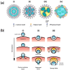
Structure and cellular entry mechanisms of lipid-based gene delivery vectors. (a) Structural diversity of lipid-based vectors for gene delivery: (i) Lamellar lipoplex: multilamellar structure with DNA/RNA sandwiched between cationic lipid bilayers. (ii) Hexagonal phase lipoplex: inverted hexagonal phase with nucleic acids enclosed within lipid-lined water channels. (iii) Solid lipid nanoparticle (SLN)/nanostructured lipid carrier (NLC): solid or solid–liquid matrix core surrounded by lipid monolayer. (iv) Nanoghost: cell membrane-derived vesicle retaining native membrane proteins and incorporating genetic cargo. (b) Pathways for cellular entry and genetic cargo delivery. (i) Direct fusion pathway: immediate fusion with plasma membrane; direct cytoplasmic release of genetic material; bypasses endosomal compartmentalization. (ii) Endosomal fusion escape: internalization via endocytosis; fusion with endosomal membrane; controlled release of genetic cargo; membrane merger preserves compartment integrity. (iii) Endosomal rupture pathway: pH-dependent ionization of lipids; osmotic pressure buildup (“proton sponge effect”); endosomal membrane destabilization and rupture; bulk release of vesicle contents.
Figure 3
Figure 3
Membrane fusion pathway and associated energy landscape. (a) Sequential stages of membrane fusion. Key feature shown in cross-sectional view. (b) Free energy profile of the fusion cascade.
Figure 4
Figure 4
Factors modulating lipid-based vector approaching cellular membrane. (a) Strategies enhancing vector-membrane adhesion. (b) Barriers impeding vector-membrane contact.
Figure 5
Figure 5
Sources of hydrophobic defect formation in lipid-based vector shell: (i) solid–liquid crystalline phase coexistence; (ii) lipid packing defects produced by inverted conical lipids; (iii) lipophilic moieties in lipid-based vector formulation.
Figure 6
Figure 6
Driving forces of membrane reorganization after its initial contact: (i) the accumulation of inverted-conical lipids (indicated in yellow) that facilitate highly curved temporary structures; (ii) lipid mixing under high chemical potential gradient blue and red arrows indicate the flux direction of lipids from different membranes, shown as red and blue circles.
Figure 7
Figure 7
Strategies for enhanced pore formation in hemifusion diaphragm: (i) diaphragm thinning by employment of cationic lipids with shorter acyl chains; (ii) ionizable lipids having cone shape in protonated state. Red and blue circles indicate lipids from two different membranes.

Similar articles

Cited by

References

    1. Hosseinkhani H., Domb A.J., Sharifzadeh G., Nahum V. Gene Therapy for Regenerative Medicine. Pharmaceutics. 2023;15:856. doi: 10.3390/pharmaceutics15030856. - DOI - PMC - PubMed
    1. Symes J.F. Gene Therapy for Ischemic Heart Disease: Therapeutic Potential. Am. J. Cardiovasc. Drugs. 2001;1:159–166. doi: 10.2165/00129784-200101030-00001. - DOI - PubMed
    1. Fus-Kujawa A., Prus P., Bajdak-Rusinek K., Teper P., Gawron K., Kowalczuk A., Sieron A.L. An Overview of Methods and Tools for Transfection of Eukaryotic Cells in Vitro. Front. Bioeng. Biotechnol. 2021;9:701031. doi: 10.3389/fbioe.2021.701031. - DOI - PMC - PubMed
    1. Tsai H.-C., Pietrobon V., Peng M., Wang S., Zhao L., Marincola F.M., Cai Q. Current Strategies Employed in the Manipulation of Gene Expression for Clinical Purposes. J. Transl. Med. 2022;20:535. doi: 10.1186/s12967-022-03747-3. - DOI - PMC - PubMed
    1. Fajrial A.K., He Q.Q., Wirusanti N.I., Slansky J.E., Ding X. A Review of Emerging Physical Transfection Methods for CRISPR/Cas9-Mediated Gene Editing. Theranostics. 2020;10:5532–5549. doi: 10.7150/thno.43465. - DOI - PMC - PubMed

LinkOut - more resources