. 2024 Dec 20;25(24):13629.
doi: 10.3390/ijms252413629.

Mucosal Bacterial Immunotherapy Attenuates the Development of Experimental Colitis by Reducing Inflammation Through the Regulation of Myeloid Cells

Affiliations

Mucosal Bacterial Immunotherapy Attenuates the Development of Experimental Colitis by Reducing Inflammation Through the Regulation of Myeloid Cells

Eva Jiménez et al. Int J Mol Sci. .

Abstract

Ulcerative colitis is a chronic relapsing-remitting and potentially progressive form of inflammatory bowel disease in which there is extensive inflammation and mucosal damage in the colon and rectum as a result of an abnormal immune response. MV130 is a mucosal-trained immunity-based vaccine used to prevent respiratory tract infections in various clinical settings. Additionally, MV130 may induce innate immune cells that acquire anti-inflammatory properties and promote tolerance, which could have important implications for chronic inflammatory diseases such as ulcerative colitis. This work demonstrated that the prophylactic administration of MV130 substantially mitigated colitis in a mouse model of acute colitis induced by dextran sulphate sodium. MV130 downregulated systemic and local inflammatory responses, maintained the integrity of the intestinal barrier by preserving the enterocyte layer and goblet cells, and reduced the oedema and fibrosis characteristic of the disease. Mechanistically, MV130 significantly reduced the infiltration of neutrophils and pro-inflammatory macrophages in the intestinal wall of the diseased animals and favoured the appearance of M2-polarised macrophages. These results suggest that MV130 might have therapeutic potential for the treatment of ulcerative colitis, reducing the risk of relapse and the progression of disease.

Keywords: MV130; bacterial immunotherapy; immunomodulation; inflammation; macrophages; neutrophils; ulcerative colitis; vaccine.


Conflict of interest statement

J.L.S. is shareholder of Inmunotek S.L. The remaining authors declare that the research was conducted in the absence of any commercial or financial relationships that could be construed as a potential conflict of interest.

Figures

Figure 1
Figure 1
Beneficial effect of prophylactic MV130 treatment on the development of acute colitis. (A) Schematic diagram of study design. Briefly, after 21 days of prophylactic MV130 treatment or vehicle, acute colitis was induced in the animals by treatment with 2.5% DSS for 7 days. (B) The disease activity index (DAI) was calculated as specified in the Materials and Methods and the mean value ± SEM for each day is plotted in the graph. Animals fed fresh water were used as controls. (C) The mean colon length ± SEM of 4 animals per group is shown. (D) Representative macroscopic image of the colon from one animal in each group. Graph shows the mean values ± SEM of the different groups of animals. Significance is indicated with respect to CTRL (*) or CTRL-DSS (#) (* p < 0.05; **** p ≤ 0.0001; # p < 0.05 and ## p < 0.01)—analysed by Kruskal–Wallis test for DAI and one-way ANOVA for colon length).
Figure 2
Figure 2
Systemic effects of MV130 in colitis-induced animals. (AJ) Mice were treated as described in Figure 1A, and samples were collected on day 4 and 7 after colitis induction. (A) Serum levels of several cytokines in the different groups of mice 7 days after DSS treatment (n = 4 per group) were measured by CBA. (B) Data represent TNFα/IL-10 ratio production in each experimental group. (C) Splenocytes were labelled with CFSE and stimulated with ConA + PMA for four days, as described in the Material and Methods. The percentage of proliferation of splenocytes from mice of different experimental groups is shown (n = 4 per group). Flow cytometry analysis of spleen (DG) and mesenteric lymph node immune populations (HJ). (D) Representative dot plots of the Treg lymphocyte population (CD4+CD25+FoxP3+) in the spleen after four days of stimulation with ConA and PMA in CTRL-DSS and MV130-DSS mice (n = 4 per group). Absolute numbers of neutrophils (E,H) and monocytes (F,I) recovered from the spleen and MLN of mice on days 4 and 7 after colitis induction (n = 3–4 per group). (G,J) Representative dot plot of myeloid populations on day 7 after DSS induction. Neutrophils (Ly6GhiLy6Clo/−) and monocyte/macrophage populations (Ly6GLy6C+) were gated within CD45+ CD11b+. (K) Flow cytometry analysis of human PBMCs after culture with MV130-CFSE for 24 h. Dot plots show the percentage of uptake of CFSE-MV130 by lymphocytes, monocytes, and granulocytes. Data are representative of 3 independent experiments. (L) mRNA expression of several cytokines was studied in monocytes treated with MV130 for 24 h by RT-qPCR. The graphs show the mean values ± SEM and the significance is indicated with respect to CTRL (*) or CTRL-DSS (#). * p < 0.05; *** p < 0.001; # p < 0.05—analysed by (A,B,E,F,H,I) Kruskal–Wallis, (C) one-way ANOVA, and (L) Mann–Whitney test.
Figure 3
Figure 3
MV130 treatment prevents colitis-induced tissue damage. (A) Haematoxylin and eosin staining was performed, and the areas of the different histological regions were measured. Graphs show the mean values ± SEM of the different groups of animals. Representative images of colon sections from each group of animals: (B) haematoxylin and eosin (scale bar: left column, 100 μm; right column, 50 μm; insert, 10 μm); (C) PAS–haematoxylin (scale bar, 100 μm); and (D) Masson’s trichrome (scale bar, 100 μm). (E) Percentage of area with loss of crypt cytoarchitecture in the mucosa. (F) Percentage of injured epithelium. (G) Quantification of goblet cells in the mucosa using PAS staining. Graphs represent mean ± SEM. The significance is indicated with respect to CTRL (*) or CTRL-DSS (#) (** p < 0.01; *** p < 0.001; # p < 0.05; ## p < 0.01—analysed by one-way ANOVA test).
Figure 4
Figure 4
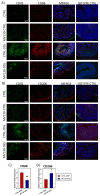
MV130 treatment reduces innate immune cell infiltration and modulates the macrophage phenotype. Colon cryosections from the different experimental groups of mice sacrificed on day 7 were immunostained for CD45 (green) and for (A) CD68 (red) or (B) CD206 (red). The isotype controls are also shown. Hoechst was used for nuclear counterstaining. Scale bars represent 100 mm. Images are representative of four animals per group. Measurement of (C) CD68+ and (D) CD206+ macrophages in the colonic mucosa and submucosa of mice from the different groups was performed. Relative CD68 or CD206 expression was calculated by dividing all individual data by the mean expression in the CTRL group of animals. Results represent the mean ± SEM of four different animals, and the significance is indicated with respect to CTRL-DSS (# p < 0.05; ## p < 0.01—analysed by Student’s t test).
Figure 5
Figure 5
MV130 induces M2-like macrophage differentiation. Human CD14+ monocytes were cultured for 6 days with GM-CSF to induce M1 macrophages or with M-CSF to induce M2 macrophages. MV130 treatment was added at the beginning of the culture. (A) Dot plots show the percentage of M2-like CD14+CD163+ cells present in the different culture conditions (representative of 4 independent experiments). (B) Percentage of macrophage viability was measured by flow cytometry. Annexin V PI cells were considered as viable cells. (CE) After 6 days of culture, supernatants were analysed for different immunomodulatory factors produced by macrophages. (C) Data represent TNFα/IL-10 ratio production, (D) IL-4, and (E) CXCL10 protein secretion by macrophages under the different experimental conditions. (F) IDO1 mRNA expression levels quantified in macrophages under the different experimental conditions at day 6 of culture. (BF) Data are mean ± SEM of four independent experiments. Significance is indicated relative to M1 control (* p < 0.05; ** p < 0.01—analysed by Kruskal–Wallis test).
Figure 6
Figure 6
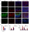
MV130 reduces the recruitment of neutrophils and tissue damage mediators in ulcerative colitis. (A) Immunodetection of CD45 (green) and CD15 (neutrophils, red) in colon cryosection 7 days after DSS induction. The isotype controls are also shown. Hoechst was used for nuclear counterstaining. Scale bars represent 100 μm. Images are representative of four animals per group. (B) Measurement of CD15+ neutrophils in the colonic mucosa and submucosa of mice from the different groups was performed (dotted line). Relative CD15 expression was calculated by dividing all individual data by the mean expression in the CTRL group. (C) Relative MPO activity in colonic protein extracts on days 4 and 7 after colitis induction. (D) Protein levels in colonic mucosa measured by flow cytometry. (BD) Results represent the mean ± SEM of four different animals per group and the significance is indicated with respect to CTRL (*) or CTRL-DSS (#). * p < 0.05; ** p < 0.01; *** p < 0.001; **** p < 0.0001; # p < 0.05; ## p < 0.01; ### p < 0.001—analysed by (B) Student’s t test, (C) Kruskal–Wallis test, (D) and one-way ANOVA test.

Similar articles

References

    1. Gros B., Kaplan G.G. Ulcerative Colitis in Adults: A Review. JAMA. 2023;330:951–965. doi: 10.1001/jama.2023.15389. - DOI - PubMed
    1. Le Berre C., Honap S., Peyrin-Biroulet L. Ulcerative colitis. Lancet. 2023;402:571–584. doi: 10.1016/S0140-6736(23)00966-2. - DOI - PubMed
    1. Shah S.C., Khalili H., Gower-Rousseau C., Olen O., Benchimol E.I., Lynge E., Nielsen K.R., Brassard P., Vutcovici M., Bitton A., et al. Sex-Based Differences in Incidence of Inflammatory Bowel Diseases-Pooled Analysis of Population-Based Studies from Western Countries. Gastroenterology. 2018;155:1079–1089.e3. doi: 10.1053/j.gastro.2018.06.043. - DOI - PubMed
    1. Saez A., Herrero-Fernandez B., Gomez-Bris R., Sánchez-Martinez H., Gonzalez-Granado J.M. Pathophysiology of Inflammatory Bowel Disease: Innate Immune System. Int. J. Mol. Sci. 2023;24:1526. doi: 10.3390/ijms24021526. - DOI - PMC - PubMed
    1. Na Y.R., Stakenborg M., Seok S.H., Matteoli G. Macrophages in intestinal inflammation and resolution: A potential therapeutic target in IBD. Nat. Rev. Gastroenterol. Hepatol. 2019;16:531–543. doi: 10.1038/s41575-019-0172-4. - DOI - PubMed

MeSH terms

Substances

LinkOut - more resources