. 2024 Dec 20;25(24):13649.
doi: 10.3390/ijms252413649.

Characterization of Site-Specific N- and O-Glycopeptides from Recombinant Spike and ACE2 Glycoproteins Using LC-MS/MS Analysis

Affiliations

Characterization of Site-Specific N- and O-Glycopeptides from Recombinant Spike and ACE2 Glycoproteins Using LC-MS/MS Analysis

Ju Hwan Song et al. Int J Mol Sci. .

Abstract

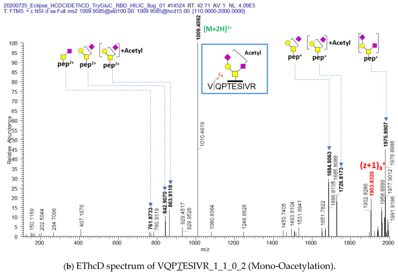
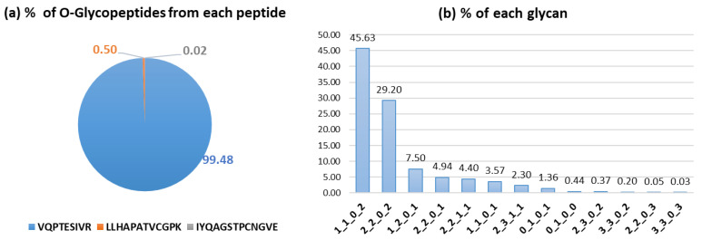
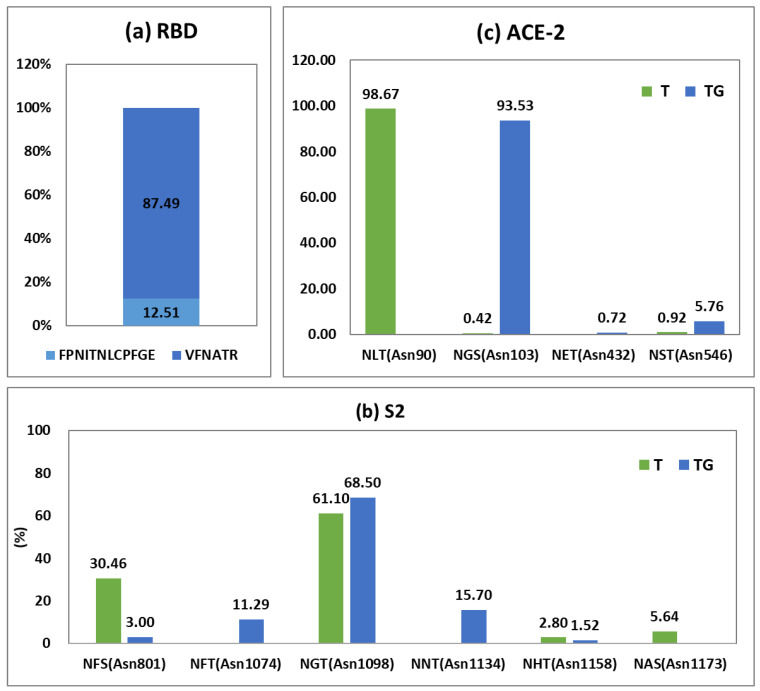
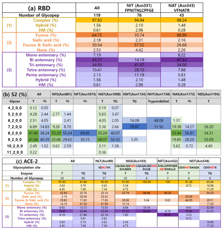
The COVID-19 pandemic, caused by severe acute respiratory syndrome coronavirus 2 (SARS-CoV-2), has resulted in hundreds of millions of infections and millions of deaths globally. Although vaccination campaigns are mitigating the pandemic, emerging viral variants continue to pose challenges. The spike (S) protein of SARS-CoV-2 plays a critical role in viral entry by binding to the angiotensin-converting enzyme 2 (ACE2) receptor, making both proteins essential targets for therapeutic and vaccine development. The glycosylation of these proteins influences their structure and function. This underscores the need for detailed site-specific glycoproteomic analysis. In this study, we characterized the N- or O-glycosylation profiles of the recombinant receptor-binding domain (RBD) of spike protein and ACE2 proteins expressed from Expi293F cells, as well as the S2 subunit of spike protein expressed in plant (N. benthamiana) cells. Using a high-resolution Orbitrap Eclipse Tribrid mass spectrometer equipped with the Ultimate 3000 RSLCnano and I-GPA (Integrated GlycoProteome Analyzer) developed in a previous study, 148 N- and 28 O-glycopeptides from RBD, 71 N-glycopeptides from the S2 subunit, and 139 N-glycopeptides from ACE2 were characterized. In addition, we report post-translational modifications (PTMs) of glycan, including mannose-6-phosphate (M6P) and GlcNAc-1-phosphate-6-O-mannose in N-glycan of RBD and ACE2, and O-acetylation in O-glycan of RBD, identified for the first time in these recombinant proteins. The relative abundance distribution according to glycosites and glycan types were analyzed by quantified site-specific N- and O (only from RBD)-glycopeptides from RBD, S2, and ACE2 using I-GPA. Asn331 for RBD, Asn1098 for S2, and Asn103 for ACE2 were majorly N-glycosylated, and dominant glycan-type was complex from RBD and ACE2 and high-mannose from S2. These findings will provide valuable insights into the glycosylation patterns that influence protein function and immunogenicity and offer new perspectives for the development of vaccines and antibody-based therapies against COVID-19.

Keywords: LC-MS/MS analysis; modification of glycan; reaction-binding domain; recombinant proteins; site-specific glycopeptides; spike glycoprotein.


Conflict of interest statement

The authors declare the following financial interest/personal relationships which may be considered as potentially competing.

Figures

Figure 1
Figure 1
EThcD spectra of (a) non-acetylated and (b) acetylated VQPTESIVR_1_1_0_2 O-glycopeptides from RBD of recombinant spike protein; 1_1_0_2 is the number of Hexose (Hex), N-acetylgalactosamine (GalNAc), fucose (Fuc), and N-acetyl neuraminic acid (NeuAc), respectively (the number of Hex_HexNac_Fuc_NeuAc). The italic and underlined T (T) or S (S) letters indicate the O-glycosylation site. The yellow circle indicates the galactose, the yellow square indicates the GalNAc, and the purple square indicates the NeuAc. The blue arrows indicate the match of theoretical m/z and actual m/z. The blue and the red colored letters indicate the c series and z (and z + 1) series ions of glycosylated peptide respectively. The green [M+2H] indicates the precursor ion.
Figure 1
Figure 1
EThcD spectra of (a) non-acetylated and (b) acetylated VQPTESIVR_1_1_0_2 O-glycopeptides from RBD of recombinant spike protein; 1_1_0_2 is the number of Hexose (Hex), N-acetylgalactosamine (GalNAc), fucose (Fuc), and N-acetyl neuraminic acid (NeuAc), respectively (the number of Hex_HexNac_Fuc_NeuAc). The italic and underlined T (T) or S (S) letters indicate the O-glycosylation site. The yellow circle indicates the galactose, the yellow square indicates the GalNAc, and the purple square indicates the NeuAc. The blue arrows indicate the match of theoretical m/z and actual m/z. The blue and the red colored letters indicate the c series and z (and z + 1) series ions of glycosylated peptide respectively. The green [M+2H] indicates the precursor ion.
Figure 2
Figure 2
HCD and CID spectra of VFNATR_7_2_0_0 and VFNATR_7_2_0_0 (+Mannose-6-Phosphate). The italic and underlined N (N) letter indicates the N-glycosylation site of peptide. 7_2_0_2 is the number of Hexose (Hex), N-acetylglucosamine (GlcNAc), fucose (Fuc), and N-acetyl neuraminic acid (NeuAc), respectively (the number of Hex_HexNac_Fuc_NeuAc). The green circle and the blue square indicate the mannose and the GlcNAc respectively. The blue arrows indicate the match of theoretical m/z and actual m/z. The blue letters indicate the y series ions. The +P means that the glycopeptide contains the mannose-6-phosphate. The red dotted boxes are the fragment ions with M6P.
Figure 3
Figure 3
HCD and CID spectra of VFNATR_6_3_0_0 and VFNATR_6_2_0_0 (+GlcNAc-1-Phosphate). The italic and underlined N (N) letter indicates the N-glycosylation site of peptide. 6_2_0_0 is the number of Hexose (Hex), N-acetylglucosamine (GlcNAc), fucose (Fuc), and N-acetyl neuraminic acid (NeuAc), respectively (the number of Hex_HexNac_Fuc_NeuAc). The green circle, the blue square and the yellow circle indicates the mannose, the GlcNAc, and the galactose respectively. The blue arrows indicate the match of theoretical m/z and actual m/z. The blue letters indicate the y series ions. The P and circled P indicates the phosphate. The red dotted boxes are related to the fragment ions with Phosphate from GlcNAc-1-Phosphate or GlcNAc-1-Phosphate. The red, green and blue boxes are the pares of the fragment ions not containing P, containing P and GlcNAc-1-Phosphate.
Figure 4
Figure 4
The quantitative percentage according to O-glycosites (a) and O-glycans (b) from RBD protein.
Figure 5
Figure 5
The quantitative distribution according to N-glycosites from (a) RBD, (b) S2, and (c) ACE-2 proteins. RBD sample was treated with trypsin and Glu-C. T: trypsin-treated S2 or ACE-2 samples. TG: trypsin- and Glu-C-treated S2 or ACE-2 samples.
Figure 6
Figure 6
The quantitative distribution according to (a) glycan type (complex, hybrid, and high-mannose (HM)), (b) fucose (containing fucose but not sialic acid), sialic acid (containing sialic acid but not fucose), fucose and sialic acid (containing fucose and sialic acid), and none (glycopeptides without fucose or sialic acid), (c) the number of branches in N-glycans for all N-glycopeptides from RBD, S2, or ACE-2 protein. The RBD sample was treated with trypsin and Glu-C. T: trypsin-treated S2 or ACE-2 samples. TG: trypsin- and Glu-C-treated S2 or ACE-2 samples. A darker color indicates a higher percentage.

Similar articles

References

    1. Walls A.C., Park Y.J., Tortorici M.A., Wall A., McGuire A.T., Veesler D. Structure, Function, and Antigenicity of the SARS-CoV-2 Spike Glycoprotein. Cell. 2020;181:281–292.e286. doi: 10.1016/j.cell.2020.02.058. - DOI - PMC - PubMed
    1. Zhang F., Schmidt F., Muecksch F., Wang Z., Gazumyan A., Nussenzweig M.C., Gaebler C., Caskey M., Hatziioannou T., Bieniasz P.D. SARS-CoV-2 spike glycosylation affects function and neutralization sensitivity. mBio. 2024;15:e0167223. doi: 10.1128/mbio.01672-23. - DOI - PMC - PubMed
    1. Zhang Y., Zhao W., Mao Y., Chen Y., Wang S., Zhong Y., Su T., Gong M., Du D., Lu X., et al. Site-specific N-glycosylation Characterization of Recombinant SARS-CoV-2 Spike Proteins. Mol. Cell. Proteom. 2021;20:100058. doi: 10.1074/mcp.RA120.002295. - DOI - PMC - PubMed
    1. Xu C., Ng D.T. Glycosylation-directed quality control of protein folding. Nat. Rev. Mol. Cell Biol. 2015;16:742–752. doi: 10.1038/nrm4073. - DOI - PubMed
    1. Jayaprakash N.G., Surolia A. Role of glycosylation in nucleating protein folding and stability. Biochem. J. 2017;474:2333–2347. doi: 10.1042/BCJ20170111. - DOI - PubMed

MeSH terms

LinkOut - more resources