. 2024 Dec 20;25(24):13662.
doi: 10.3390/ijms252413662.

Different Cytotoxic Effects of Cisplatin on Pancreatic Ductal Adenocarcinoma Cell Lines

Affiliations

Different Cytotoxic Effects of Cisplatin on Pancreatic Ductal Adenocarcinoma Cell Lines

Antonella Muscella et al. Int J Mol Sci. .

Abstract

This study examined the response to cisplatin in BxPC-3, Mia-Paca-2, PANC-1, and YAPC pancreatic cancer lines with different genotypic and phenotypic characteristics, and the mechanisms associated with their resistance. BxPC-3 and MIA-PaCa-2 cell lines were the most sensitive to cisplatin, while YAPC and PANC-1 were more resistant. Consistently, in cisplatin-treated BxPC-3 cells, the cleavage patterns of pro-caspase-9, -7, -3, and PARP-1 demonstrated that they were more sensitive than YAPC cells. The autophagic pathway, promoting cisplatin resistance, was active in BxPC-3 cells, as demonstrated by the time-dependent conversion of LC3-I to LC3-II, whereas it was not activated in YAPC cells. In cisplatin-treated BxPC-3 cells, Bcl-2 decreased, while Beclin-1, Atg-3, and Atg-5 increased along with JNK1/2 phosphorylation. Basal levels of phosphorylated ERK1/2 in each cell line were positively correlated with cisplatin IC50 values, and cisplatin caused the activation of ERK1/2 in BxPC-3 and YAPC cells. Furthermore, ERK1/2 pharmacological inactivation increased cisplatin lethality in both BxPC-3 and YAPC cells, suggesting that p-ERK1/2 may be related to cisplatin resistance of PDAC cells. Different mechanisms and strategies are generally required to acquire drug resistance. Here, we partially explain the other response to cisplatin of BxPC-3 and YAPC cell lines by relating it to the role of ERK pathway.

Keywords: apoptosis; cisplatin; cytotoxicity; pancreatic ductal adenocarcinoma; signal transduction.


Conflict of interest statement

The authors declare no conflicts of interest.

Figures

Figure 1
Figure 1
Cytotoxic effects of cisplatin on pancreatic tumor lines. BxPC-3 (A), Mia Paca-2 (B), PANC-1 (C) and YAPC (D) cells were treated with different concentration of cisplatin (0.1–200 µM). Cell viability was measured with sulforhodamine B (SRB) colorimetric assay, after 12, 24, 48, or 72 h. Data are the means ± standard deviation (SD) of five independent experiments with eight replicates in each and are presented as percent of control at the corresponding time point, with the control set as 100% ** p < 0.01; *** p < 0.001 by one-way ANOVA followed by Bonferroni/Dunn post hoc tests. The dashed lines indicate the IC50 values.
Figure 2
Figure 2
Analysis of mitochondrial membrane potential using the cationic dye JC-1 in BxPC-3 (A) and (B) YAPC cells. (A) Cells were incubated, or not, with 50 μM cisplatin for 24 hours and stained with 4′,6-diamidino-2-phenylindole (DAPI). The representative fields by confocal microscopy (magnification 40×) of one of four independent experiments are shown. (B) Quantification of the percentage of apoptotic nuclei was obtained using DAPI (means ± SD; n = 5). For both BxPC-3 and YAPC cells: p < 0.001 by one-way ANOVA followed by Bonferroni/Dunn post hoc tests; values with shared letters are not significantly different according to Bonferroni/Dunn. (C,D) Fluorescent spectra of JC-1 in BxPC-3 and YAPC cells treated or not with 50 μM cisplatin for the indicated time. The data are means ± S.D. of five different experiments and are presented as red J-aggregates/green monomer JC-1 fluorescence ratio. Asterisks indicate values that are significantly different (p < 0.05) from control at the same time point.
Figure 3
Figure 3
The cleavage of caspase-9, caspase-7, caspase-3 and Parp-1 induced by cisplatin in BxPC-3 (A) and YAPC (B) cells. Cells were treated with 50 µM cisplatin for the indicated time, and then subjected to Western blotting. Incubation with anti-β-actin confirmed the equal protein loading. The results shown are representative of five different experiments. The histograms on the right are representative of five independent experiments and the densitometry results are expressed as the mean ± SD (n = 5) of the sum of the gray level values of the westerns. (A) p < 0.001 by one-way ANOVA for all proteins. (B) p < 0.001 by one-way ANOVA for caspase-9 and -7; p > 0.05 by one-way ANOVA for caspase-3 and PARP-1. The values of histograms for full-length caspases and PARP with shared lower-case letters are not significantly different according to Bonferroni/Dunn post hoc tests. The values of histograms for cleaved caspases and PARP with shared capital case letters are not significantly different according to Bonferroni/Dunn post hoc tests.
Figure 4
Figure 4
Cisplatin Induces Autophagy in BxPC-3 Cells. BxPC-3 (A) and YAPC (B) cells were treated with 50 µM cisplatin for different times. Cell lysates were analyzed using Western blotting, using specific antibodies. Sequential incubation with anti-β-actin confirmed the equal protein loading. Representative immunoblots of five experiments are depicted. The histograms on the right are representative of five independent experiments and the densitometry results are expressed as the mean ± SD (n = 5) of the sum of the gray level values of the westerns. (A) p < 0.001 by one-way ANOVA for all proteins. (B) p < 0.001 by one-way ANOVA for Atg3, Atg7, p-JNK1/2 and BCL2; p < 0.01 by one-way ANOVA for Beclin-1 and p > 0.05 by one-way ANOVA for LC3 I. The values of histograms with shared lower-case letters are not significantly different according to Bonferroni/Dunn post hoc tests. The values of histograms for LC3 II with shared capital case letters are not significantly different according to Bonferroni/Dunn post hoc tests.
Figure 5
Figure 5
The effects of cisplatin on ERK1/2 activation. (A) BxPC-3 and YAPC cells were treated with 50 µM cisplatin for different times and cell lysates were analyzed by Western blotting, using activated ERK1/2 specific antibody. (B) Cells were pre-treated for 45 min with 25 µM PD98059 and then incubated or not with 50 µM cisplatin for 24 h. Then, cell viability was measured with sulforhodamine B colorimetric assay. (C) Basal expression levels of ERK1/2 in cell lines. Cell lysates were analyzed using Western blotting, using anti-phospho-ERK1/2 (p-ERK1/2) or anti-total ERK1/2 antibodies. (D) The relationship between the levels of phosphorylated ERK1/2 and the IC50 of cisplatin in the pancreatic cell lines used. Sequential incubation with anti-β-actin confirmed the equal protein loading. Representative immunoblots of five experiments are depicted. The histograms on the right are representative of five independent experiments and the densitometry results are expressed as the mean ± SD (n = 5) of the sum of the gray level values of the westerns. A and B, p < 0.001 by one-way ANOVA followed by Bonferroni/Dunn post hoc tests. Values with shared letters are not significantly different according to Bonferroni/Dunn. The lower-case letters refer to BxPC-3 cells and capital case letters to YAPC cells.
Figure 6
Figure 6
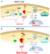
The schematic diagram illustrates the intracellular signaling mechanisms in pancreatic cancer cells. (A) BxPC-3 cells, in response to cisplatin, activate key pathways such as apoptosis (pro-caspase-9, -7, -3 cleavage), as well as autophagy (LC3-I to LC3-II conversion) and ERK1/2 signaling, promoting survival. (B) YAPC cells with a KRAS mutation exhibit constitutive ERK1/2 pathway activation; consequently, there is a decreased activation pattern of caspase-9, caspase-7, and caspase-3, along with PARP-1 cleavage, representing apoptosis resistance. Additionally, YAPC cells do not show autophagy activation.

Similar articles

Cited by

References

    1. Sung H., Ferlay J., Siegel R.L., Laversanne M., Soerjomataram I., Jemal A., Bray F. Global Cancer Statistics 2020: GLOBOCAN Estimates of Incidence and Mortality Worldwide for 36 Cancers in 185 Countries. CA Cancer J. Clin. 2021;71:209–249. doi: 10.3322/caac.21660. - DOI - PubMed
    1. Grossberg A.J., Chu L.C., Deig C.R., Fishman E.K., Hwang W.L., Maitra A., Marks D.L., Mehta A., Nabavizadeh N., Simeone D.M., et al. Multidisciplinary standards of care and recent progress in pancreatic ductal adenocarcinoma. CA Cancer J. Clin. 2020;70:375–403. doi: 10.3322/caac.21626. - DOI - PMC - PubMed
    1. Neoptolemos J.P., Cunningham D., Friess H., Bassi C., Stocken D.D., Tait D.M., Dunn J.A., Dervenis C., Lacaine F., Hickey H., et al. Adjuvant therapy in pancreatic cancer: Historical and current perspectives. Ann. Oncol. 2003;14:675–692. doi: 10.1093/annonc/mdg207. - DOI - PubMed
    1. Yokoyama Y., Nimura Y., Nagino M. Advances in the treatment of pancreatic cancer: Limitations of surgery and evaluation of new therapeutic strategies. Surg. Today. 2009;39:466–475. doi: 10.1007/s00595-008-3904-6. - DOI - PubMed
    1. Xu B., Shen W., Liu X., Zhang T., Ren J., Fan Y., Xu J. Oridonin inhibits BxPC-3 cell growth through cell apoptosis. Acta Biochim. Biophys. Sin. 2015;47:164–173. doi: 10.1093/abbs/gmu134. - DOI - PubMed

MeSH terms