. 2024 Dec 21;25(24):13673.
doi: 10.3390/ijms252413673.

LPS Disrupts Endometrial Receptivity by Inhibiting STAT1 Phosphorylation in Sheep

Affiliations

LPS Disrupts Endometrial Receptivity by Inhibiting STAT1 Phosphorylation in Sheep

Xing Fan et al. Int J Mol Sci. .

Abstract

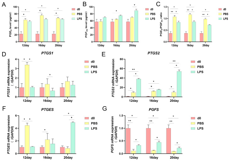
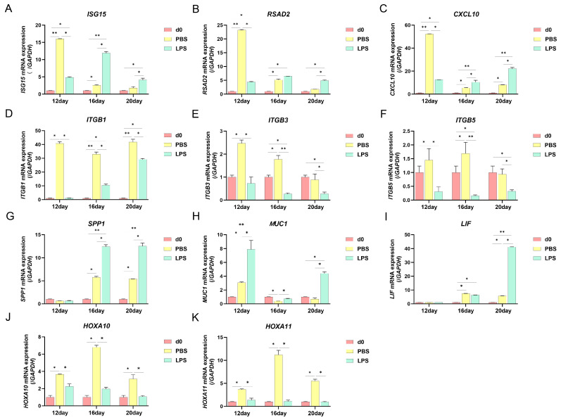
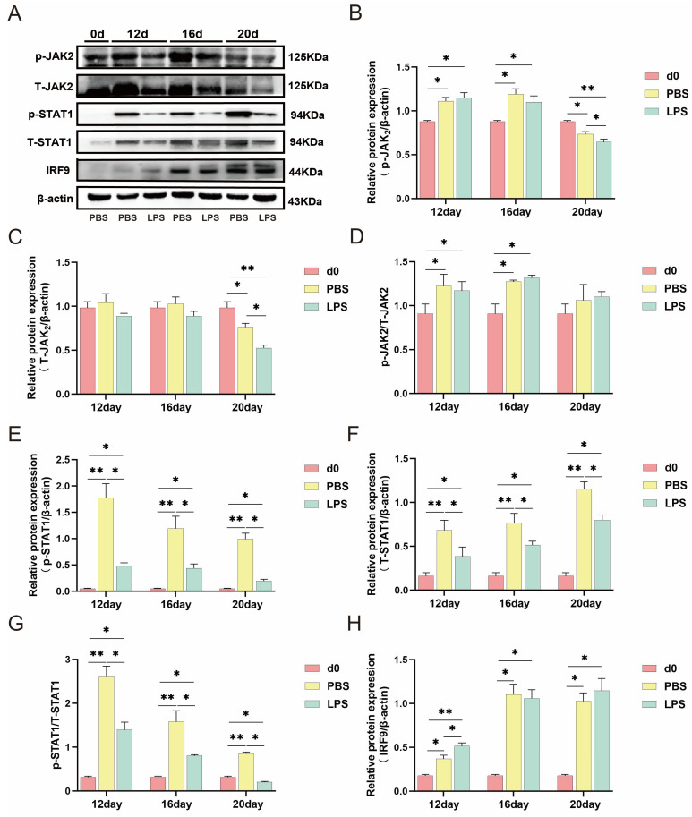
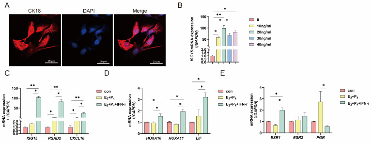
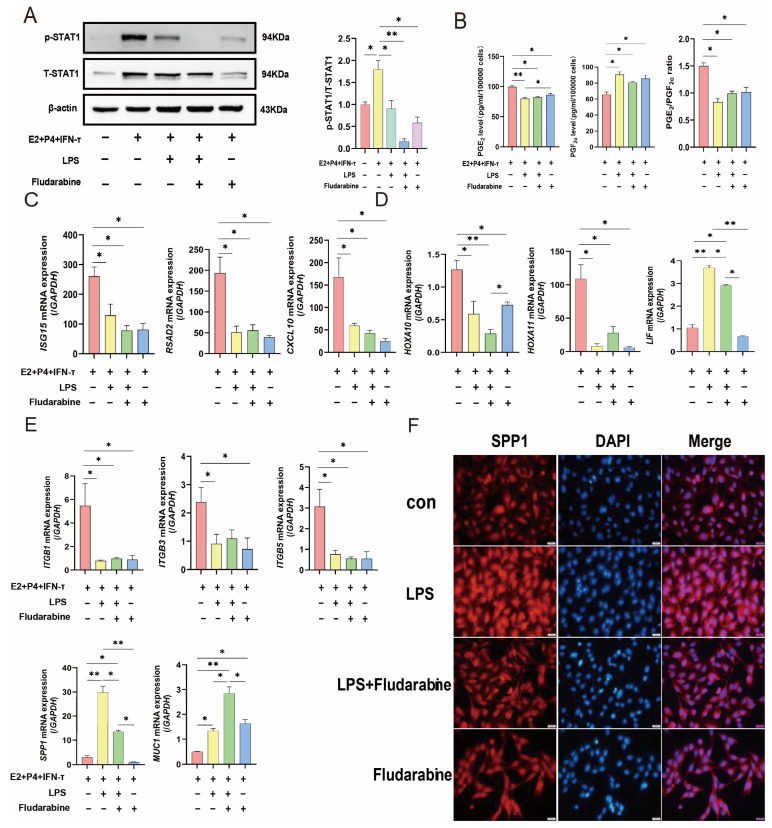
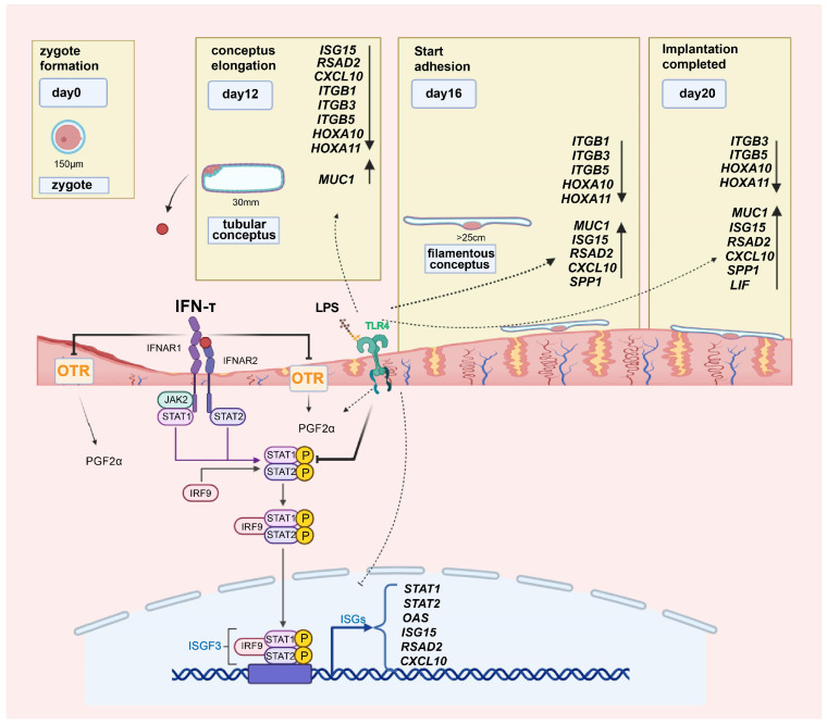
Uterine infections reduce ruminant reproductive efficiency. Reproductive dysfunction caused by infusion of Gram-negative bacteria is characterized by the failure of embryo implantation and reduced conception rates. Lipopolysaccharide (LPS), a major component of the outer membrane of Gram-negative bacteria, is highly abortogenic. In this study, the effects of LPS infusion on the endometrial receptivity of sheep were studied during three critical periods of embryo implantation. The results showed that LPS infusion on d12, d16, and d20 of pregnancy in vivo interfered with the expression of prostaglandins (PGs) and affected the expression of adhesion-related factors (ITGB1/3/5, SPP1), key implantation genes (HOXA10, HOXA11 and LIF), and progestational elongation genes (ISG15, RSAD2 and CXCL10) during embryo implantation. In addition, after LPS infusion on d12, d16, and d20, the phosphorylation level of STAT1 significantly decreased and the protein expression level of IRF9 significantly increased on d12, suggesting that LPS infusion in sheep impairs endometrial receptivity through the JAK2/STAT1 pathway. Sheep endometrial epithelial cells were treated with 17 β-estrogen, progesterone, and/or interferon-tau in vitro to mimic the receptivity of the endometrium during early pregnancy for validation. LPS and the p-STAT1 inhibitor fludarabine were both added to the model, which resulted in reduced p-STAT1 protein expression, significant inhibition of PGE2/PGF2α, and significant suppression of the expression of key embryo implantation genes. Collectively, these results indicate that LPS infusion in sheep on d12, d16, and d20 impairs endometrial receptivity through the JAK2/STAT1 pathway, which is responsible for LPS-associated pregnancy failure.

Keywords: LPS; STAT1; embryo implantation; endometrial receptivity; sheep.


Conflict of interest statement

The authors declare no conflict of interest.

Figures

Figure 1
Figure 1
Effect of LPS on prostaglandin expression in sheep endometrium. (A) The secretion of PGE2 and PGF2α in endometrial tissue was measured on d12, d16, and d20 of pregnancy using an ELISA kit. (B) The secretion of PGE2 and PGF2α in endometrial tissue was measured on d12, d16, and d20 of pregnancy using an ELISA kit. (C) The ratio of PGE2 and PGF2α in endometrial tissue on d12, d16, and d20 of pregnancy. (D) The rate-limiting enzymes PTGS1, PTGS2 (E), PTGES (F), and PGFS (G) of synthesized PGs in endometrial tissue on d12, d16, and d20 of pregnancy were measured by real-time quantitative PCR. All data are presented as the mean ± SEM, n ≥ 3; * p < 0.05; ** p < 0.01.
Figure 2
Figure 2
Effect of LPS on endometrial receptivity genes in sheep. (A) The pro-conceptus elongation gene ISG15, RSAD2 (B), and CXCL10 (C) on d12, d16, and d20 of pregnancy in endometrial tissue were measured by real-time quantitative PCR. (D) The adhesion molecules ITGB1, ITGB3 (E), ITGB5 (F), SPP1 (G), and MUC1 (H) on d12 of pregnancy in endometrial tissue were measured by real-time quantitative PCR. (I) The endometrial receptivity markers HOXA10, HOXA11 (J), and LIF (K) on d12 of pregnancy in endometrial tissue were measured by real-time quantitative PCR. All data are presented as the mean ± SEM, n ≥ 3; * p < 0.05; ** p < 0.01.
Figure 3
Figure 3
LPS affected JAK2/STAT1 pathways. (A) The protein level of p-JAK2, T-JAK2, p-STAT1, T-STAT1, and IRF9 on d12, d16, and d20 of pregnancy in sheep endometrial tissue. (B) p-JAK2/β-actin, T-JAK2/β-actin (C), and p-JAK2/T-JAK2 (D) ratio on d12, d16, and d20 of pregnancy in sheep endometrial tissue. (E) p-STAT1/β-actin, T-STAT1/β-actin (F), and p-STAT1/T-STAT1 (G) ratio on d12, d16, and d20 of pregnancy in sheep endometrial tissue. (H) The IRF9/β-actin ratio on d12, d16, and d20 of pregnancy in sheep endometrial tissue. All data are presented as the mean ± SEM, n ≥ 3; * p < 0.05; ** p < 0.01.
Figure 4
Figure 4
Establishment of a receptive sheep endometrial epithelial cell model for sheep. (A) Confocal microscopy was used to observe the morphology of sEECs. Red: Cy3-labeled cytokeratin 18 protein; blue, DAPI-labeled nuclei; scale bar: 20 µm. (B) Expression of ISG15 was measured under different concentrations in sEECs. (CE) The endometrial receptivity-related genes ISG15, RSAD2, CXCL10, HOXA10, HOXA11, LIF, ESR1, ESR2, and PGR in sEECs were measured by real-time quantitative PCR. GAPDH (sheep) was used as the reference gene in all samples. sEECs: sheep endometrial epithelial cells. All data are presented as the mean ± SEM, n ≥ 3; * p < 0.05; ** p < 0.01.
Figure 5
Figure 5
Effect of LPS or fludarabine treatment on the expression of endometrial receptivity-related genes under hormone treatment. (A) The protein level of p-STAT1 and T-STAT1 in sEECs. (B) The secretion of PGE2 and PGF2α in sEECs. (CE) The pro-conceptus elongation genes ISG15, RSAD2, CXCL10, adhesion molecules ITGB1/3/5, MUC1, SPP1, and receptivity markers HOXA10, HOXA11, LIF mRNA expression levels in sEECs. GAPDH (sheep) was used as the reference gene in all samples. (F) Confocal microscope images of SPP1 expression in four treatment groups. Red: Cy3-labeled SPP1 protein; blue, DAPI-labeled nuclei; scale bar: 20 µm. All data are presented as the mean ± SEM, n ≥ 3; * p < 0.05; ** p < 0.01.
Figure 6
Figure 6
Schematic characterization of the cellular mechanism of LPS infusion effects on endometrial receptivity in sheep during early pregnancy. LPS blocked the effect of IFN-τ in the three stages of sheep embryo implantation and impaired the endometrial receptivity, which is characterized by interfering with the secretion of prostaglandins, hindering the elongation of the conceptus, and reducing the adhesion of the embryo by inhibiting the phosphorylation of STAT1.

Similar articles

Cited by

References

    1. Yang Q., Liu J., Wang Y., Zhao W., Wang W., Cui J., Yang J., Yue Y., Zhang S., Chu M., et al. A proteomic atlas of ligand–receptor interactions at the ovine maternal–fetal interface reveals the role of histone lactylation in uterine remodeling. J. Biol. Chem. 2021;298:101456. doi: 10.1016/j.jbc.2021.101456. - DOI - PMC - PubMed
    1. Chen Q., Zhang A., Yu F., Gao J., Liu Y., Yu C., Zhou H., Xu C. Label-free proteomics uncovers energy metabolism and focal adhesion regulations responsive for endometrium receptivity. J. Proteome Res. 2015;14:1831–1842. doi: 10.1021/acs.jproteome.5b00038. - DOI - PubMed
    1. Spencer T.E., Johnson G.A., Bazer F.W., Burghardt R. Implantation mechanisms: Insights from the sheep. Reproduction. 2004;128:657–668. doi: 10.1530/rep.1.00398. - DOI - PubMed
    1. Hue I., Degrelle S.A., Turenne N. Conceptus elongation in cattle: Genes, models and questions. Anim. Reprod. Sci. 2012;134:19–28. doi: 10.1016/j.anireprosci.2012.08.007. - DOI - PubMed
    1. Spencer T., Johnson G., Bazer F., Burghardt R. Fetal-maternal interactions during the establishment of pregnancy in ruminants. Reprod. Domest. Rumin. 2007;64:379–396. doi: 10.5661/RDR-VI-379. - DOI - PubMed

LinkOut - more resources