Review
. 2024 Dec 21;25(24):13681.
doi: 10.3390/ijms252413681.

Somatic Instability Leading to Mosaicism in Fragile X Syndrome and Associated Disorders: Complex Mechanisms, Diagnostics, and Clinical Relevance

Affiliations
Review

Somatic Instability Leading to Mosaicism in Fragile X Syndrome and Associated Disorders: Complex Mechanisms, Diagnostics, and Clinical Relevance

Dragana Protic et al. Int J Mol Sci. .

Abstract

Fragile X syndrome (FXS) is a genetic condition caused by the inheritance of alleles with >200 CGG repeats in the 5' UTR of the fragile X messenger ribonucleoprotein 1 (FMR1) gene. These full mutation (FM) alleles are associated with DNA methylation and gene silencing, which result in intellectual disabilities, developmental delays, and social and behavioral issues. Mosaicism for both the size of the CGG repeat tract and the extent of its methylation is commonly observed in individuals with the FM. Mosaicism has also been reported in carriers of premutation (PM) alleles, which have 55-200 CGG repeats. PM alleles confer risk for the fragile X premutation-associated conditions (FXPAC), including FXTAS, FXPOI, and FXAND, conditions thought to be due to the toxic consequences of transcripts containing large CGG-tracts. Unmethylated FM (UFM) alleles are transcriptionally and translationally active. Thus, they produce transcripts with toxic effects. These transcripts do produce some FMRP, the encoded product of the FMR1 gene, albeit with reduced translational efficiency. As a result, mosaicism can result in a complex clinical presentation. Here, we review the concept of mosaicism in both FXS and in PM carriers, including its potential clinical significance.

Keywords: FMR1 gene; activation ratio; full mutation; methylation; mosaicism; premutation.


Conflict of interest statement

The authors declare no conflicts of interest.

Figures

Figure 4
Figure 4
Activation ratio. Southern blot analyses (panels a,b) and capillary electropherograms (c) of PCR products (triplet repeat PCR with/without the CGG primer and methylation PCR) from three females with a premutation: one with a complete skewed AR (line1), and the other two with an AR of 0.41 and 0.38, respectively (lines 2 and 3). One female with a full mutation and a very skewed AR of approximately 0.85 is shown in lane 4. Measurements of methylation status and activation ratio (AR) are shown as previously described [81]. For Southern blot analysis, DNA was digested with EcoRI/NruI and probed as described in [65]. MW = 1 kb size ladder marker; Cntrl F = normal female, negative control. M = normal male. In the electropherograms plots, the x-axis indicates the number of base pairs, and the y-axis indicates the relative fluorescence intensity.
Figure 1
Figure 1
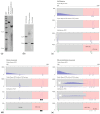
Representative examples of FMR1 mosaic categories. Southern blot analysis (a,b) shows the presence of non-methylated and methylated FMR1 alleles in DNA isolated from peripheral blood derived from three males: full mutation, 100% methylated (a, line 2); full mutation size mosaic (b, line 2); and full mutation size/methylation mosaic (a, line 3). 1 kb molecular weight size marker (MW, a line 1); female and male negative controls (b, lines 1 and 3). The normal unmethylated (2.8 kb) and a normal methylated (5.2 kb) allele size are shown on the left (b). Southern blot analysis was carried out on genomic DNA digested with EcoRI and NruI and probed with the specific FMR1 genomic probe StB12.3, as described in [65]. Corresponding electropherograms of triplet repeat PCR with/without the CGG primer and methylation PCR from peripheral blood DNA of (c) a full mutation male [(a) Southern blot lane 2], of (d) a full mutation size mosaic male [(b) Southern blot lane 2], and (e) a full mutation size/methylation mosaic male [(a) Southern blot lane 3] are shown. Southern blot and PCR analysis of a male with a full mutation (c) show the presence of a methylated FM greater than 200 CGG repeats. Southern blot analysis of a full mutation size mosaic male (d) shows PM fragments above 5.2 kb, indicating the presence of several FMR1 methylated alleles greater than 200 CGG in size and of an allele in the premutation range. Triplet repeats PCR analysis with/without the CGG primer electropherograms shows the presence of a premutation allele with 100 CGG and FM (>200 CGG) alleles. mPCR reveals that the PM allele of 100 CGG was unmethylated, and the FM alleles were completely methylated. Southern blot analysis of a full mutation size/methylation mosaic male (e) shows the presence of a continuum of unmethylated alleles ranging approximately from 90 to 750 CGG repeats (~70% unmethylation) and the presence of hypermethylated alleles greater than 200 CGG repeats (above 5.2 kb, 30% methylation). The triplet repeats PCR with/without the CGG primer and mPCR electropherograms show the presence of a broad range of unmethylated alleles ranging from the normal to the low full mutation range. PCR and methylation PCR were carried out as reported in [66,67]. In the electropherogram plots, the x-axis indicates the number of base pairs, and the y-axis indicates the relative fluorescence intensity. Methylation percentage was calculated as previously described in [68].
Figure 2
Figure 2
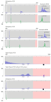
Inter- and intra-tissue mosaicism. Southern blot analyses (SB; a,d) and electropherograms of PCR products (triplet repeat PCR with/without the CGG primer and methylation PCR) from DNA isolated from peripheral blood (panels b,e) and primary fibroblast cells (panels c,f) of two examples of FM mosaic males (examples 1 and 2) are shown. Both SB and PCR analyses show CGG instability, as illustrated by the presence of highly heterogeneous fragments across all amplification ranges and differences in FMR1 methylation when comparing blood to primary cultured fibroblasts. For SB analysis, DNA was digested with EcoRI and NruI and probed as described in [65]. MW = 1 kb size ladder marker; Cntr Female = normal female control. Male FM = full mutation male, positive control. In the electropherogram plots, the x-axis indicates the number of base pairs, and the y-axis indicates the relative fluorescence intensity. Methylation percentage was calculated as described in [68].
Figure 3
Figure 3
Somatic allelic instability in PM carriers. Panels (a,b) show examples of methylation PCR profiles observed in two female PM carriers. Capillary electropherograms show increasing levels of somatic expansion. (c) Example of triplet repeats PCR with/without the CGG primer and mPCR in a male with somatic instability (FM size/methylation mosaic), characterized by the presence of FM alleles and a wide range of premutation alleles with a complete absence of methylation. The x-axis indicates the number of base pairs, and the y-axis indicates relative fluorescence intensity.

Similar articles

References

    1. Brown C.J., Robinson W.P. The causes and consequences of random and non-random X chromosome inactivation in humans. Clin. Genet. 2000;58:353–363. doi: 10.1034/j.1399-0004.2000.580504.x. - DOI - PubMed
    1. Paulson H. Repeat expansion diseases. Handb. Clin. Neurol. 2018;147:105–123. doi: 10.1016/b978-0-444-63233-3.00009-9. - DOI - PMC - PubMed
    1. Wright G.E.B., Collins J.A., Kay C., McDonald C., Dolzhenko E., Xia Q., Bečanović K., Drögemöller B.I., Semaka A., Nguyen C.M., et al. Length of Uninterrupted CAG, Independent of Polyglutamine Size, Results in Increased Somatic Instability, Hastening Onset of Huntington Disease. Am. J. Hum. Genet. 2019;104:1116–1126. doi: 10.1016/j.ajhg.2019.04.007. - DOI - PMC - PubMed
    1. Lee H.G., Imaichi S., Kraeutler E., Aguilar R., Lee Y.W., Sheridan S.D., Lee J.T. Site-specific R-loops induce CGG repeat contraction and fragile X gene reactivation. Cell. 2023;186:2593–2609.e2518. doi: 10.1016/j.cell.2023.04.035. - DOI - PMC - PubMed
    1. Sidorov M.S., Auerbach B.D., Bear M.F. Fragile X mental retardation protein and synaptic plasticity. Mol. Brain. 2013;6:15. doi: 10.1186/1756-6606-6-15. - DOI - PMC - PubMed

Substances