

EWSR1::ATF1 Translocation: A Common Tumor Driver of Distinct Human Neoplasms
- PMID: 39769457
- PMCID: PMC11728112
- DOI: 10.3390/ijms252413693
EWSR1::ATF1 Translocation: A Common Tumor Driver of Distinct Human Neoplasms
Abstract
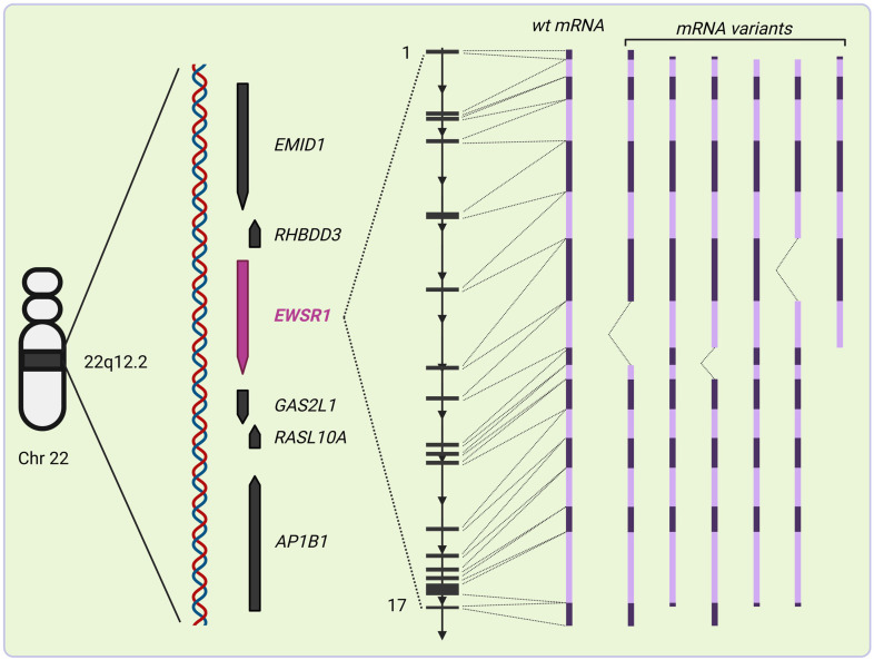
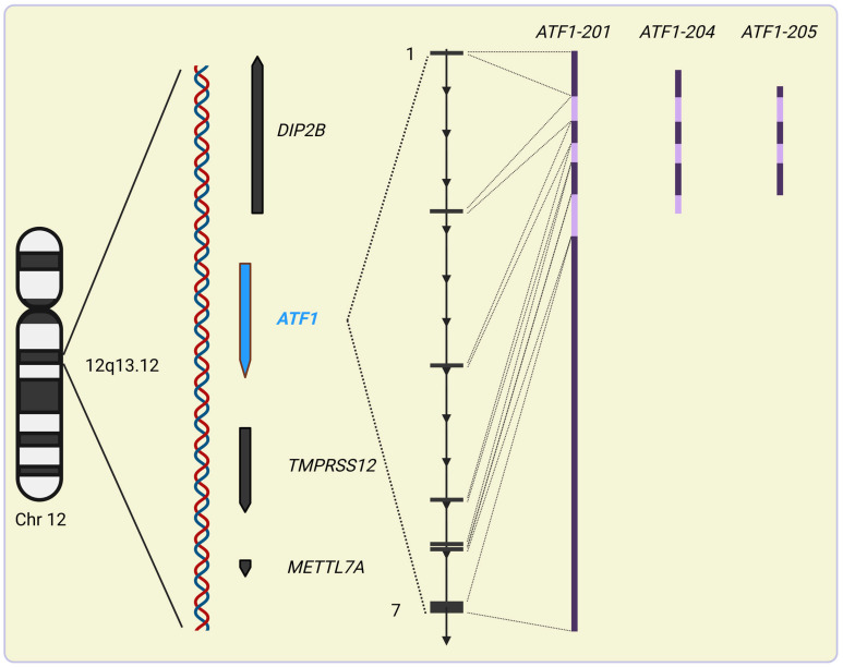
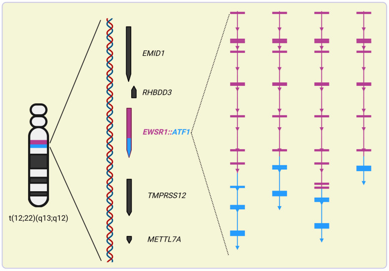
Cancer is among the leading causes of mortality in developed countries due to limited available therapeutic modalities and high rate of morbidity. Although malignancies might show individual genetic landscapes, recurring aberrations in the neoplastic genome have been identified in the wide range of transformed cells. These include translocations of frequently affected loci of the human genetic material like the Ewing sarcoma breakpoint region 1 (EWSR1) of chromosome 22 that results in malignancies with mesodermal origin. These cytogenetic defects frequently result in the genesis of fusion genes involving EWSR1 and a number of genes from partner loci. One of these chromosomal rearrangements is the reciprocal translocation between the q13 and q12 loci of chromosome 12 and 22, respectively, that is believed to initiate cancer formation by the genesis of a novel, chimeric transcription factor provoking dysregulated gene expression. Since soft-tissue neoplasms carrying t(12;22)(q13;q12) have very poor prognosis and clinical modalities specifically targeting t(12;22)(q13;q12)-harboring cells are not available to date, understanding this DNA aberration is not only timely but urgent. Here, we review our current knowledge of human malignancies carrying the specific subset of EWSR1 rearrangements that leads to the expression of the EWSR1::ATF1 tumor-driver chimeric protein.
Keywords: Ewing sarcoma region 1; activating transcription factor 1; chimeric proteins; gene fusion; malignant mesothelioma; reciprocal chromosome translocation.
Conflict of interest statement
J.R.B., Y.L. and A.P. declare no conflicts of interest. Z.F. is a shareholder and scientific adviser of Translocon Biothechnologies PLC, Budapest, Hungary.
Figures

Similar articles
-
Protein arginine methyltransferase 5 is essential for oncogene product EWSR1-ATF1-mediated gene transcription in clear cell sarcoma.J Biol Chem. 2022 Oct;298(10):102434. doi: 10.1016/j.jbc.2022.102434. Epub 2022 Aug 27. J Biol Chem. 2022. PMID: 36041632 Free PMC article.
-
Detection of specific gene rearrangements by fluorescence in situ hybridization in 16 cases of clear cell sarcoma of soft tissue and 6 cases of clear cell sarcoma-like gastrointestinal tumor.Diagn Pathol. 2018 Sep 15;13(1):73. doi: 10.1186/s13000-018-0752-6. Diagn Pathol. 2018. PMID: 30219084 Free PMC article.
-
RNA sequencing identifies fusion of the EWSR1 and YY1 genes in mesothelioma with t(14;22)(q32;q12).Genes Chromosomes Cancer. 2013 Aug;52(8):733-40. doi: 10.1002/gcc.22068. Epub 2013 Apr 30. Genes Chromosomes Cancer. 2013. PMID: 23630070
-
Complex rearrangement of chromosomes 19, 21, and 22 in Ewing sarcoma involving a novel reciprocal inversion-insertion mechanism of EWS-ERG fusion gene formation: a case analysis and literature review.Cancer Genet Cytogenet. 2008 Mar;181(2):81-92. doi: 10.1016/j.cancergencyto.2007.11.002. Cancer Genet Cytogenet. 2008. PMID: 18295659 Review.
-
Identification of EWSR1 rearrangements in patients with immature hematopoietic neoplasms: A case series and review of literature.Ann Diagn Pathol. 2022 Jun;58:151942. doi: 10.1016/j.anndiagpath.2022.151942. Epub 2022 Mar 23. Ann Diagn Pathol. 2022. PMID: 35344861 Review.
References
-
- Nassour J., Martien S., Martin N., Deruy E., Tomellini E., Malaquin N., Bouali F., Sabatier L., Wernert N., Pinte S., et al. Defective DNA single-strand break repair is responsible for senescence and neoplastic escape of epithelial cells. Nat. Commun. 2016;7:10399. doi: 10.1038/ncomms10399. - DOI - PMC - PubMed
Publication types
MeSH terms
Substances
LinkOut - more resources
Full Text Sources
Medical
