. 2024 Dec 22;25(24):13705.
doi: 10.3390/ijms252413705.

Methodological Assessment of ExoGAG for Isolation of Cerebrospinal Fluid Extracellular Vesicles as a Source of Biomarkers

Affiliations

Methodological Assessment of ExoGAG for Isolation of Cerebrospinal Fluid Extracellular Vesicles as a Source of Biomarkers

Nil Salvat-Rovira et al. Int J Mol Sci. .

Abstract

Extracellular vesicles (EVs) in cerebrospinal fluid (CSF) represent a valuable source of biomarkers for central nervous system (CNS) diseases, offering new pathways for diagnosis and monitoring. However, existing methods for isolating EVs from CSF often prove to be labor-intensive and reliant on specialized equipment, hindering their clinical application. In this study, we present a novel, clinically compatible method for isolating EVs from CSF. We optimized the use of ExoGAG, a commercially available reagent that has been tested in plasma, urine and semen, and compared it directly with differential ultracentrifugation using Western blotting, protein quantification, nanoparticle tracking analysis, and cryogenic electron microscopy. Additionally, we analyzed the presence of specific microRNAs (miRNAs) known to be present in CSF-derived EVs. Our data demonstrate that ExoGAG is an effective method for isolating EVs from CSF, yielding a higher amount of EVs compared to traditional ultracentrifugation methods, and with comparable levels of specific miRNAs. In conclusion, the use of ExoGAG in a clinical setting may facilitate the testing of biomarkers essential for tracking brain pathology in CNS diseases.

Keywords: ExoGAG; biomarkers; cerebrospinal fluid; extracellular vesicles; isolation method; miRNAs; ultracentrifugation.


Conflict of interest statement

The authors declare no conflict of interest.

Figures

Figure 1
Figure 1
Effectiveness of ExoGAG in isolating EVs from CSF. (a) Schematic representation of the method used to precipitate EVs with ExoGAG. SN = supernatant. (b) Representative immunoblots of CD81 and the intracellular markers GM130 and calnexin, in EVs isolated with ExoGAG using different sample-to-reagent (CSF/ExoGAG) ratios. A longer exposure of calnexin is shown. Quantification of (c) CD81 and (d) CD81/calnexin ratio. CD81/calnexin levels were normalized to the average of the 1:2 ratio. N = 6 independent CSF samples. Data are represented as mean ± S.E.M. (e) Representative immunoblots of CD81 and Alix as EV markers, along with GM130 and calnexin in EVs isolated with ExoGAG when using the 2:1 sample-to-reagent ratio. Cell lysate was used as positive control (+C). Imaging of the stain-free membrane is shown for total protein in both Western blot panels.
Figure 2
Figure 2
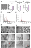
Comparative analysis of ExoGAG and UC isolated CSF EVs: protein estimation, NTA, and Cryo-EM. Quantification of total protein estimated by (a) Sypro staining or (b) BCA in EVs isolated with ExoGAG or UC. N = 3 for Sypro and N = 4 for BCA of independent CSF samples. Data are represented as mean ± S.E.M. (c) Comparison of the total particle (EVs) concentration after ExoGAG or UC isolation estimated by NTA. N = 3 independent CSF samples. Data are represented as mean ± S.E.M. * p < 0.05; ** p < 0.01 by Student’s t-test. Number and size profiling of EVs isolated with (d) ExoGAG or (e) UC by NTA. N = 3 independent EV samples per technique, each with three technical replicates (except for sample 1-UC, which only had two technical replicates). Data are represented as mean ± S.E.M. (shown as red lines). Cryo-EM images at different magnifications showing the presence of vesicles (indicated by red arrows) after using (f) ExoGAG at a 3:1 sample-to-reagent ratio or (g) UC. Calibration bars are indicated at the bottom of each image.
Figure 3
Figure 3
EV enrichment in ExoGAG-isolated versus UC-isolated EVs. (a) Representative immunoblots of EVs isolated using ExoGAG or UC, along with corresponding supernatants (Sn) and starting CSF. The absence of the intracellular marker calnexin in the EV preparations is also shown. Cell lysate (+C) was used as a positive control. The total amount of protein is shown in the stain-free gel and in the Sypro-stained membrane. (b) Quantification of CD81 and Alix levels in EVs isolated with ExoGAG or UC, based on N = 4 independent CSF samples. Data are represented as mean ± S.E.M. * p < 0.05 by Student’s t-test.
Figure 4
Figure 4
Presence of specific miRNAs in CSF EVs isolated by UC or ExoGAG. (a) Threshold cycle (Ct) values for the spike-in UniSp2, UniSp4, and UniSp6 in EVs isolated by UC and ExoGAG. (b) Delta Ct values for the determinations of miR-204a-5p and let7a-5p in EVs isolated by UC and ExoGAG. Data are represented as mean ± S.E.M., based on N = 5 independent CSF samples.

References

    1. Perez-Gonzalez R., Gauthier S.A., Kumar A., Levy E. The exosome secretory pathway transports amyloid precursor protein carboxyl-terminal fragments from the cell into the brain extracellular space. J. Biol. Chem. 2012;287:43108–43115. doi: 10.1074/jbc.M112.404467. - DOI - PMC - PubMed
    1. Hook V., Podvin S., Mosier C., Boyarko B., Seyffert L., Stringer H., Rissman R.A. Emerging evidence for dysregulated proteome cargoes of tau-propagating extracellular vesicles driven by familial mutations of tau and presenilin. Extracell. Vesicles Circ. Nucleic Acids. 2023;4:588–598. doi: 10.20517/evcna.2023.44. - DOI - PMC - PubMed
    1. Jeon I., Cicchetti F., Cisbani G., Lee S., Li E., Bae J., Lee N., Li L., Im W., Kim M., et al. Human-to-mouse prion-like propagation of mutant huntingtin protein. Acta Neuropathol. 2016;132:577–592. doi: 10.1007/s00401-016-1582-9. - DOI - PMC - PubMed
    1. Howitt J., Hill A.F. Exosomes in the Pathology of Neurodegenerative Diseases. J. Biol. Chem. 2016;291:26589–26597. doi: 10.1074/jbc.R116.757955. - DOI - PMC - PubMed
    1. van Niel G., D’Angelo G., Raposo G. Shedding light on the cell biology of extracellular vesicles. Nat. Rev. Mol. Cell Biol. 2018;19:213–228. doi: 10.1038/nrm.2017.125. - DOI - PubMed

MeSH terms

LinkOut - more resources