. 2024 Dec 22;25(24):13715.
doi: 10.3390/ijms252413715.

MET Exon 14 Skipping and Novel Actionable Variants: Diagnostic and Therapeutic Implications in Latin American Non-Small-Cell Lung Cancer Patients

Affiliations

MET Exon 14 Skipping and Novel Actionable Variants: Diagnostic and Therapeutic Implications in Latin American Non-Small-Cell Lung Cancer Patients

Solange Rivas et al. Int J Mol Sci. .

Abstract

Targeted therapy indications for actionable variants in non-small-cell lung cancer (NSCLC) have primarily been studied in Caucasian populations, with limited data on Latin American patients. This study utilized a 52-genes next-generation sequencing (NGS) panel to analyze 1560 tumor biopsies from NSCLC patients in Chile, Brazil, and Peru. The RNA sequencing reads and DNA coverage were correlated to improve the detection of the actionable MET exon 14 skipping variant (METex14). The pathogenicity of MET variants of uncertain significance (VUSs) was assessed using bioinformatic methods, based on their predicted driver potential. The effects of the predicted drivers VUS T992I and H1094Y on c-MET signaling activation, proliferation, and migration were evaluated in HEK293T, BEAS-2B, and H1993 cell lines. Subsequently, c-Met inhibitors were tested in 2D and 3D cell cultures, and drug affinity was determined using 3D structure simulations. The prevalence of MET variants in the South American cohort was 8%, and RNA-based diagnosis detected 27% more cases of METex14 than DNA-based methods. Notably, 20% of METex14 cases with RNA reads below the detection threshold were confirmed using DNA analysis. The novel actionable T992I and H1094Y variants induced proliferation and migration through c-Met/Akt signaling. Both variants showed sensitivity to crizotinib and savolitinib, but the H1094Y variant exhibited reduced sensitivity to capmatinib. These findings highlight the importance of RNA-based METex14 diagnosis and reveal the drug sensitivity profiles of novel actionable MET variants from an understudied patient population.

Keywords: MET exon 14 skipping; c-Met inhibitors; next-generation sequencing; non-small-cell lung cancer; novel actionable variants.


Conflict of interest statement

R.A. received honoraria for conferences, advisory boards, and educational activities from Roche and Janssen, as well as grants and support for scientific research from Pfizer, Roche, and Thermo Fischer Scientific. The founders had no role in the study’s design; in the collection, analysis, or interpretation of data; the writing of the manuscript; or the decision to publish the results.

Figures
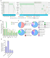
Figure 1
Figure 1
The mutational profiles of NSCLC actionable genes in South America evidenced a high prevalence of MET variants. (A) Each column of the oncoplots represents a patient, and the rows show the prevalence of the variants in the eight NSCLC actionable genes. (B) Comparison of variant prevalence in eight actionable NSCLC genes. (C) The gender, subject country, tumor stage, and tobacco use information of the patients with variants of the MET gene. (D) MET variant categorization according to the clinical significance, the number, and the percentage of the patients. * p-value ≤ 0.05; ** p-Value ≤ 0.01.
Figure 2
Figure 2
The RNA and DNA MET sequencing analysis evidenced differences in diagnosing the MET exon 14 skipping variant. (A) DNA regions of exons 13, 14, 15, and introns 14 and 15 of the MET gene (GRCh37.p13). Below are the DNA variants’ locations, which affect the coding sequence of exon 14 and the splicing donor region of the MET gene (variants located in red in rows) of each patient with a DNA variant in the SD region. (B) The broad spectrum of the RNA reads for the METex14 variant is shown in the x-axis. Each column represents a patient; the dashedline shows the threshold (120 reads) for the positive METex14 diagnosis [26]. (C) The number of patients categorized as negative and positive for METex14, according to the numbers of RNA reads. (D) The conceptual map represents all TBx from the NSCLC patients with a pair of RNA- and DNA-sequenced QC pass data. (E) The Pearson correlation between the RNA and DNA reads is represented by a continous line and the standard error as a shadow. (F) The Pearson correlation analysis between the allele frequency of the positive METex14 DNA variants (X-axis) and the number of RNA reads (Y-axis). (G) Altered genes in the tumor profile of the patients with low RNA reads for METex14.
Figure 3
Figure 3
T992I and H1094Y were the most prevalent and bioinformatically predicted drivers and actionable. (A) All the VUSs were localized in the Met protein domains. The green, red, blue, and yellow rectangles represent the location of the Sema, PSI, TIG, and kinase protein domains, respectively. Above the lolliplot, (I) blue dots represent regions sensitive to targeted therapies, according to Oncokb. (II) The exons are represented by blue and light-blue boxes. (III-IV-V) The subcellular location of the mature protein. (B) The driver prediction of the VUSs located at the JM and TK domains (x-axis) using the bioinformatic algorithms CGI, Cadd13, polyphen2, mutation taster, and sift. Light pink and white represent predicted passengers and tolerated variants; green represents those variants’ predicted drivers.
Figure 4
Figure 4
The VUSs predicted to be drivers, T992I and H1094Y, promote the survival of proliferative non-tumor cells and migration in tumor cells. (A) Representative Western blots of total Met and β-actin expression, evaluated for the H1993 GFP (basal), METex14, T992I, and H1094Y cells. (B) Densitometry levels of total normalized Met/β-actin (+SEM). The graph represents the normalized average from 3 independent experiments, ±SEM. (C) The representative Western blots of total Met and β-actin expression were evaluated for the HEK293T GFP (basal), METex14, T992I, and H1094Y cells. (D) Densitometry levels of total normalized Met/β-actin (+SEM). The graph represents the normalized average from 3 independent experiments, ±SEM. (E) The absorbance averages of HEK293T and H1993 cells expressing GFP, METex14, T992I, and H1094Y; the cells incubated with and without HGF. (F) Representative microphotographies of the wound healing at 0 and 24 h of H1993 cells expressing METex14, T992I, and H1094Y, treated with and without HGF were taken at 4×. (G) The wound closure percentage was calculated for each experimental condition. Finally, three independent experiments averaging the ±SEM are shown. A two-way ANOVA with Tukey correction was applied, and the p-values were adjusted for multiple comparisons. * p < 0.05; ** p < 0.01; *** p < 0.001; **** p < 0.0001; n.s. non-significant.
Figure 5
Figure 5
The VUSs predicted to be drivers, T992I and the H1094Y, increased Met-activating phosphorylation and the downstream Akt signaling pathway. (A) Representative Western blot images of total Met, total Akt, β-actin, Met p(Y1230-1234-1235), and Akt p(S473) protein expression of HEK293T cells. (B,C) The densitometry levels of Met phosphorylation, Akt phosphorylation, and β-actin were normalized relative to the total Met and Akt. (D) Representative Western blots of total Met, total Akt, β-actin, Met p(Y1230-1234-1235), and Akt p(S473) protein expression of H1993 cells. (E,F) Densitometry levels of Met phosphorylation, Akt phosphorylation, and β-actin normalized relative to the total Met and Akt for H1993 cells. Graphs represent the normalized average from 3 independent experiments, ±SEM. (G) Densitometry levels of Metp, relative to β-actin levels. (H) Pearson correlation between the Metp and Met total, relative to β-actin levels. A one-way ANOVA with Tukey correction was applied, and the p-values were adjusted for multiple comparisons. * p < 0.05; ** p < 0.01; *** p < 0.001; n.s. non-significant.
Figure 6
Figure 6
The 2D and 3D cell cultures expressing the Met-predicted driver variants were sensitive to c-Met inhibitors. (A) A total of 2000 HEK293T-expressing variants were seeded in 2D and incubated for 24 h with crizotinib, capmatinib, and savolitinib. The absorbance was calculated from three independent experiments and normalized, relative to the non-treatment culture cells. (B) Representative microphotographs (taken at 10×) were captured with a Cytation3 imaging reader of the 3D H1993 cells (spheroid) on each day of their life. On day 2 of spheroid formation (~200 µm sphere diameter), the drugs were incubated, and then the cells were released from the treatment until day 5. (C) The spheroids were treated with savolitinib for 24 h. Each experimental condition consisted of triplicates, averaged for each experimental condition. The three independent experiments were averaged, ±SEM. * p < 0.05; ** p < 0.01; *** p < 0.001; **** p < 0.0001 and n.s non-significant.
Figure 7
Figure 7
Molecular dynamics simulations illustrate the savolitinib–MET protein system binding at an approximate mean distance of 3.0. The stability of the pyridazinone ring of savolitinib within the binding site of the MET WT:SLB (A), MET H1094Y:SLB (B), and MET T992I:SLB (C) complexes is largely determined by hydrophobic interactions. Importantly, within the METex14:SLB complex (D), savolitinib is incapable of achieving a stable conformation due to substantial modifications in the initial loop that precedes the tyrosine kinase domain. Therefore, an unstable pocket site was produced. To clarify its proximity to savolitinib, the H1094 residue in the METex14:SLB complex (D) is highlighted in yellow in this context.

Similar articles

Cited by

References

    1. Hammerschmidt S., Wirtz H. Lung cancer: Current diagnosis and treatment. Dtsch. Arztebl. Int. 2009;106:809–818; quiz 819–820. doi: 10.3238/arztebl.2009.0809. - DOI - PMC - PubMed
    1. Passiglia F., Bertolaccini L., Del Re M., Facchinetti F., Ferrara R., Franchina T., Malapelle U., Menis J., Passaro A., Pilotto S., et al. Diagnosis and treatment of early and locally advanced non-small-cell lung cancer: The 2019 AIOM (Italian Association of Medical Oncology) clinical practice guidelines. Crit. Rev. Oncol. Hematol. 2020;148:102862. doi: 10.1016/j.critrevonc.2019.102862. - DOI - PubMed
    1. Polanco D., Pinilla L., Gracia-Lavedan E., Mas A., Bertran S., Fierro G., Seminario A., Gomez S., Barbe F. Prognostic value of symptoms at lung cancer diagnosis: A three-year observational study. J. Thorac. Dis. 2021;13:1485–1494. doi: 10.21037/jtd-20-3075. - DOI - PMC - PubMed
    1. Sharma A., Jasrotia S., Kumar A. Effects of Chemotherapy on the Immune System: Implications for Cancer Treatment and Patient Outcomes. Naunyn. Schmiedebergs. Arch. Pharmacol. 2023;397:2551–2566. doi: 10.1007/s00210-023-02781-2. - DOI - PubMed
    1. Howlader N., Forjaz G., Mooradian M.J., Meza R., Kong C.Y., Cronin K.A., Mariotto A.B., Lowy D.R., Feuer E.J. The Effect of Advances in Lung-Cancer Treatment on Population Mortality. N. Engl. J. Med. 2020;383:640–649. doi: 10.1056/NEJMoa1916623. - DOI - PMC - PubMed

MeSH terms

LinkOut - more resources