Review
. 2024 Dec 23;25(24):13727.
doi: 10.3390/ijms252413727.

The Impact of Calcium Overload on Cellular Processes: Exploring Calcicoptosis and Its Therapeutic Potential in Cancer

Affiliations
Review

The Impact of Calcium Overload on Cellular Processes: Exploring Calcicoptosis and Its Therapeutic Potential in Cancer

Adrianna Gielecińska et al. Int J Mol Sci. .

Abstract

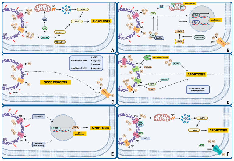
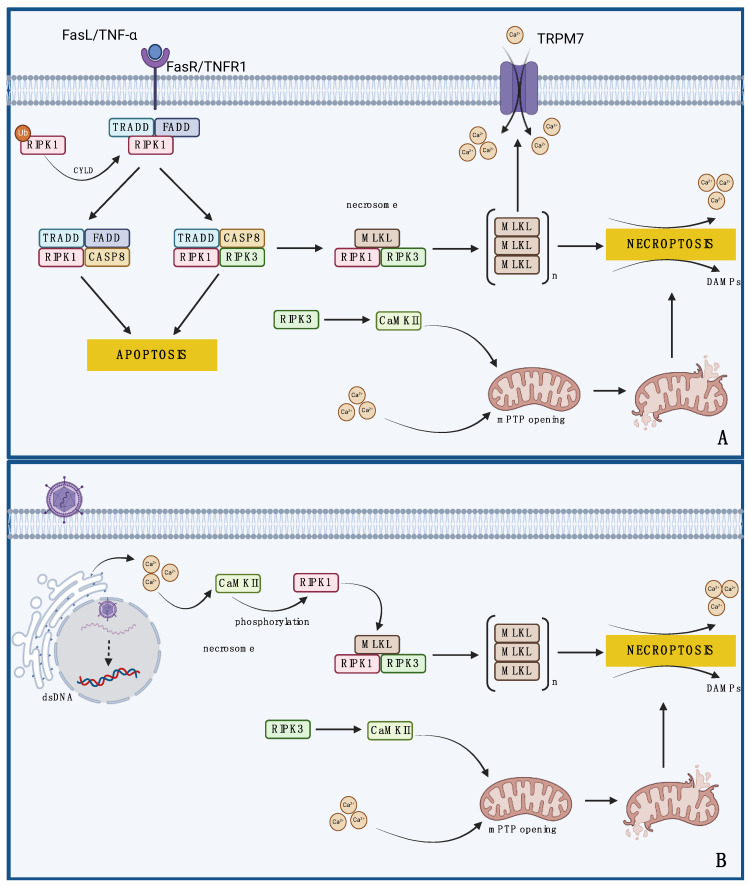
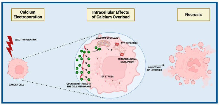
The key role of calcium in various physiological and pathological processes includes its involvement in various forms of regulated cell death (RCD). The concept of 'calcicoptosis' has been introduced as a calcium-induced phenomenon associated with oxidative stress and cellular damage. However, its definition remains controversial within the research community, with some considering it a general form of calcium overload stress, while others view it as a tumor-specific calcium-induced cell death. This review examines 'calcicoptosis' in the context of established RCD mechanisms such as apoptosis, necroptosis, ferroptosis, and others. It further analyzes the intricate relationship between calcium dysregulation and oxidative stress, emphasizing that while calcium overload often triggers cell death, it may not represent an entirely new type of RCD but rather an extension of known pathways. The purpose of this paper is to discuss the implications of this perspective for cancer therapy focusing on calcium-based nanoparticles. By investigating the connections between calcium dynamics and cell death pathways, this review contributes to the advancement of our understanding of calcicoptosis and its possible therapeutic uses.

Keywords: apoptosis; calcicoptosis; calcium; cancer; necroptosis; necrosis.


Conflict of interest statement

The authors declare no conflicts of interest.

Figures

Figure 1
Figure 1
Calcium signaling mechanisms leading to apoptosis. (A) Calcium is released into the cytoplasm through inositol triphosphate receptors (IP3Rs) and ryanodine receptors (RyRs) present in the endoplasmic reticulum (ER) membrane in response to ER stress. Free calcium ions interact with calpain, leading to the activation of caspase 12 and subsequently caspase 3, resulting in apoptosis through an ER-related pathway. Calpain also triggers mitochondrial apoptosis by affecting pro-apoptotic proteins Bcl-2 associated X protein (BAX) and BH3 interacting domain death agonist (BID), leading to the release of cytochrome C and the activation of the initiator caspase 9. (B) Calcium bound to calmodulin modulates the action of protein phosphatase (PP), which, by influencing the pro-apoptotic protein Bcl2-associated agonist of cell death (BAD), leads to the neutralization of the action of the anti-apoptotic protein BCL-2. Calmodulin is transported to the nucleus by the γ subunit of calcium/calmodulin-stimulated protein kinase II (CaMKIIγ) to phosphorylate transcription factors, including the cAMP-responsive element binding protein (CREB). Furthermore, calcium influx caused by the opening of calcium release-activated calcium channel protein 1 (ORAI1) causes dephosphorylation of nuclear factor of activated T cells 1 (NFAT1) by calcineurin and subsequently leads to its translocation to the nucleus, where it mediates gene expression. (C) In the ER membrane, the stromal interaction molecule 1 (STIM1) protein initiates the opening of ORAI1 channels in the plasma membrane, regulates calcium influx, and participates in the store-operated calcium entry (SOCE) process. (D) Regulation of calcium homeostasis through the inhibitor of apoptosis-stimulating protein P53 (iASPP)–transmembrane and Coiled-Coil 1 (TMCO1) axis: iASPP competes with the E3 ligase Gp78 to binding to TMCO1, preventing cell apoptosis. (E) ER stress affects the function of the C/EBP homology protein (CHOP) pathway, resulting in the expression of ER oxidase 1 alpha (ERO1-α), which increases the activity of IP3R calcium channels, increasing the release of calcium into the cytoplasm. (F) The binding of the anti-apoptotic protein BCL-2 to IP3Rs prevents the release of calcium into the cytoplasm. However, the pro-apoptotic proteins BAX and BAD, by acting on calcium channels, promote calcium release, which, in excess, leads to cell death by apoptosis via the mitochondrial pathway. BCL-2 may also interact with other calcium channels, including plasma membrane calcium ATPases (PMCAs). Created with BioRender.com (accessed on 24 October 2024).
Figure 2
Figure 2
(A) Death-receptor-mediated necroptosis: Binding of Fas ligand (FasL) or tumor necrosis factor α (TNF-α) to Fas receptor (FasR) or tumor necrosis factor receptor 1 (TNFR1) causes the assembly of the TNFR1-associated death domain protein (TRADD)/FAS-associated death domain protein (FADD)/receptor-interacting protein kinase (RIPK)-1 complex, where RIPK1 is deubiquitinated by cylindromatosis (CYLD). In the presence of caspase 8 (CASP8), the complex induces apoptotic death. In the absence of CASP8, the complex transforms into a necrosome composed of RIPK1, RIPK3, and lineage kinase domain-like protein (MLKL), leading to necroptosis. During necroptosis, damage-associated molecular patterns (DAMPs) and calcium ions are released, further driving necroptosis. MLKL trimers are translocated to the plasma membrane, where they interact with the transient receptor potential melastatin 7 (TRPM7) channel to promote calcium influx into cells. Excess calcium ions and activated calcium/calmodulin-stimulated protein kinase II (CaMKII) affect the opening of permeability transition pores (PTPs) in mitochondria, leading to their functional and structural degradation and driving necroptosis. (B) Death-receptor-independent necroptosis: As a result of the action of factors other than death receptors, e.g., viral infection, there is an increase in the concentration of cytosolic calcium, which activates CaMKII. Kinase phosphorylates RIPK1, leading to necrosome formation and cell death. In addition, the influence of calcium ions and CaMKII can also lead to the opening of PTP channels in the mitochondrial membrane and their structural and functional degradation, enhancing necroptosis. Created with BioRender.com (accessed on 24 October 2024).
Figure 3
Figure 3
Mechanism and effects of calcium electroporation in cancer therapy. Electroporation increases the permeability of the cell membrane, leading to the opening of pores that allow calcium ions to enter the cell. Normally maintained at a low level inside the cell, calcium ions rapidly increase in concentration. Mitochondria begin to accumulate excess calcium, leading to their overload and subsequent dysfunction. The mitochondrial membrane potential is disrupted, and the mitochondria lose their ability to produce adenosine triphosphate (ATP). Excess calcium also results in the overproduction of reactive oxygen species (ROS), which damage proteins, lipids, and DNA, thereby increasing oxidative stress. The loss of ATP, combined with ROS-induced damage, leads to the destruction of cellular structures and the initiation of necrosis. Created with BioRender.com (accessed on 24 October 2024).

Similar articles

Cited by

References

    1. Li L., Xin J., Wang H., Wang Y., Peng W., Sun N., Huang H., Zhou Y., Liu X., Lin Y., et al. Fluoride Disrupts Intestinal Epithelial Tight Junction Integrity through Intracellular Calcium-Mediated RhoA/ROCK Signaling and Myosin Light Chain Kinase. Ecotoxicol. Environ. Saf. 2023;257:114940. doi: 10.1016/j.ecoenv.2023.114940. - DOI - PubMed
    1. Sun Y.-B., Lou F., Irving M. Calcium- and Myosin-Dependent Changes in Troponin Structure During Activation of Heart Muscle. J. Physiol. 2009;587:155–163. doi: 10.1113/jphysiol.2008.164707. - DOI - PMC - PubMed
    1. Jordan M.R., Lopez R.A., Morrisonponce D. StatPearls. StatPearls Publishing; Treasure Island, FL, USA: 2024. Asystole. - PubMed
    1. Gilbert M., Jung S.-R., Reed B.J., Sweet I.R. Islet Oxygen Consumption and Insulin Secretion Tightly Coupled to Calcium Derived from L-Type Calcium Channels but Not from the Endoplasmic Reticulum. J. Biol. Chem. 2008;283:24334–24342. doi: 10.1074/jbc.M802097200. - DOI - PMC - PubMed
    1. Shlisky J., Mandlik R., Askari S., Abrams S., Belizan J.M., Bourassa M.W., Cormick G., Driller-Colangelo A., Gomes F., Khadilkar A., et al. Calcium Deficiency Worldwide: Prevalence of Inadequate Intakes and Associated Health Outcomes. Ann. N. Y. Acad. Sci. 2022;1512:10–28. doi: 10.1111/nyas.14758. - DOI - PMC - PubMed

LinkOut - more resources