. 2024 Dec 17;29(24):5949.
doi: 10.3390/molecules29245949.

Sphaeropsidin A Loaded in Liposomes to Reduce Its Cytotoxicity and Preserve Antifungal Activity Against Candida auris

Affiliations

Sphaeropsidin A Loaded in Liposomes to Reduce Its Cytotoxicity and Preserve Antifungal Activity Against Candida auris

Annalisa Buonanno et al. Molecules. .

Abstract

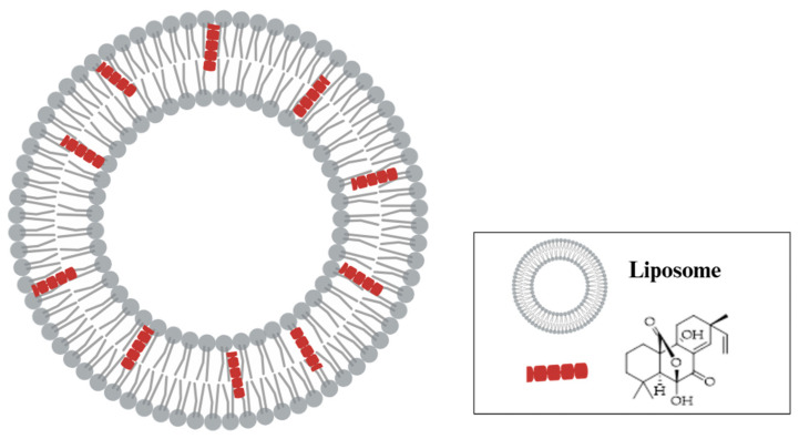
Candida species constitute the most common cause of fungal infections in humans; the emergence of resistance and biofilm formation by Candida species further threaten the limited availability of antifungal agents. Over the past decade, C. auris has caused significant outbreaks worldwide and has emerged as a human pathogenic fungus that causes diseases ranging from superficial to life-threatening disseminated infections. Despite the recent advances in antifungal research, the mechanisms of drug resistance in C. auris remain poorly understood even as its ability to form biofilms poses a significant therapeutic challenge. The purpose of this research was to elucidate the fungal properties of Sphaeropsidin A (SphA), a secondary metabolite derived from Diplodia fungi, with a specific focus on its efficacy against C. auris. This study revealed that SphA and its liposomal encapsulated (SphA-L) form are fungistatic with time-kill kinetics highlighting their efficacy and significantly inhibited the formation of C. auris biofilms. Our investigation into the antifungal mechanism of this drug revealed notable alterations in ROS production and the disruption of the Candida cell cycle. Our findings show that SphA-L impairs key pathogenic traits of C. auris, such as its ability to adhere to human epithelial cell lines, while exhibiting no harmful effects on human cells, highlighting its potential as a future therapeutic agent. In Caenorhabditis elegans infection models, both ShpA and SphA-L displayed effective antifungal activity, significantly reducing the C. auris fungal load and improving nematode survival rates, underscoring their promise as antifungal candidates. Overall, the potent antifungal effects of SphA and SphA-L against C. auris encourage further research.

Keywords: Caenorhabditis elegans; Candida auris; ROS production; Sphaeropsidin A; antifungal activity; biofilm; loaded liposome; nosocomial infection.


Conflict of interest statement

The authors declare no conflicts of interest.

Figures

Figure 1
Figure 1
Schematic representation of liposomes loaded with SphA. The figure was created with Biorender.com (License: Academic Individual Plan × 3 yrs (27 February 2023–27 February 2026)).
Figure 2
Figure 2
Time to kill assay of SphA and SphA-L on C. auris at concentrations of 17.5 μg mL−1 (1/2 MIC) and 35 μg mL−1 (1 MIC) of SphA and 25 μg mL−1 (1/2 MIC) and 50 μg mL−1 (1 MIC) of SphA-L. Data reported are the means of three independent experiments ± SDs.
Figure 3
Figure 3
Efficacy of SphA and SphA-L on C. auris at different stages of biofilm development. The sensitivity of C. auris biofilm was reported as viable cell number (%) (bar graph) and metabolic activity (line graph). SphA and SphA-L were added to attached cells and biofilms monitored following a 24 h period (A) or added to cells in a 24 h biofilm and monitored following an additional 24 h growth period (B). Data from CFU and XTT assays are represented as percent differences relative to untreated biofilm cells. Data reported are the means of three independent experiments ± SDs.
Figure 4
Figure 4
FACS flow cytometry analysis of C. auris by using propidium iodide staining (PI). (A) Flow cytometry analysis showed the DNA contents at the indicated time points of C. auris treated or not with SphA and SphA-L. Cell cycle analysis was obtained with the BD Accury C6 flow cytometer. The data were analyzed with the 10.6 version of FlowJo Program. (B) Histogram represents the mean percentages of at least three independent experiments in each cell cycle phase of untreated control and SphA and SphA-L treated cells at 0, 60, 90, 120, and 150 min.
Figure 5
Figure 5
Susceptibility of C. auris to SphA and SphA-L induced intracellular ROS production. ROS production was assessed by cell staining with H2DCFDA. Data represents the means ± SDs of three independent experiments; statistical significant is indicated by * p < 0.05 (Tukey’s test).
Figure 6
Figure 6
Cytotoxic effects of SphA and SphA-L on HaCaT cells, measured by MTT assay. Various concentrations of SphA and SphA-L (5 μg mL−1, 10 μg mL−1, 20 μg mL−1, 50 μg mL−1, and 100 μg mL−1) showed dose-dependent responses after a 24 h incubation period. Cell viability was measured at OD595 with means ± SDs (n = 3), and statistical significance is indicated by * p < 0.05, denoting significance when compared to the untreated cells (Tukey’s test).
Figure 7
Figure 7
Evaluation of the anti-adhesion effect of SphA-L on C. auris cells using HaCaT cells that were either pre-treated or post-treated. C. auris infection of HaCaT cells without treatment served as the positive control. Results represent the means of three independent experiments, with error bars indicating standard deviation. Values with dissimilar letters were significantly different from each other (a, b, c) (p < 0.05, Tukey’s test).
Figure 8
Figure 8
In vivo evaluation of the antifungal efficacy of SphA and SphA-L in C. elegans. (A) Pilot screen of C. elegans survival in the presence of SphA and SphA-L. The liquid assay assessed the survival of infected and non-infected nematodes over a 48 h period. Asterisks (*) indicate statistically significant differences between treatments, p < 0.05. (B) Survival rates of C. elegans infected with C. auris. This figure illustrates the comparative survival of nematodes exposed to E. coli OP50 as a negative control and those infected with C. auris. The survival rates were statistically different, p < 0.05. (C) Antifungal effects of SphA and SphA-L on C. elegans. The survival of infected and non-infected nematodes was evaluated in the presence of SphA and SphA-L over 48 h. Asterisks (*) indicate statistically significant differences of SphA-L compared the other treatments, p < 0.05. (D) Average number of offspring produced per nematode in various treatment groups. This figure presents the reproductive outputs of both infected and non-infected nematodes treated with SphA and SphA-L as well as controls. Asterisks (*) indicate statistically significant differences between treatments and the control group (OP50), p < 0.05.

Similar articles

Cited by

References

    1. Wanda C.R. An overview of the antimicrobial resistance mechanisms of bacteria. AIMS Microbiol. 2018;4:482–501. - PMC - PubMed
    1. Jamal M., Ahmad W., Andleeb S., Jalil F., Imran M., Nawaz M.A., Hussain T., Ali M., Rafiq M., Kamil M.A. Bacterial biofilm and associated infections. J. Chin. Med. Assoc. 2018;81:7–11. doi: 10.1016/j.jcma.2017.07.012. - DOI - PubMed
    1. Fanning S., Mitchell A.P. Fungal Biofilms. PLOS Pathog. 2012;8:e1002585. doi: 10.1371/journal.ppat.1002585. - DOI - PMC - PubMed
    1. Butassi E., Svetaz L., Carpinella M.C., Efferth T., Zacchino S. Fungal Biofilms as a Valuable Target for the Discovery of Natural Products That Cope with the Resistance of Medically Important Fungi—Latest Findings. Antibiotics. 2021;10:1053. doi: 10.3390/antibiotics10091053. - DOI - PMC - PubMed
    1. Du H., Bing J., Hu T., Ennis C.L., Nobile C.J., Huang G. Candida auris: Epidemiology, biology, antifungal resistance, and virulence. PLOS Pathog. 2020;16:e1008921. doi: 10.1371/journal.ppat.1008921. - DOI - PMC - PubMed

LinkOut - more resources